in ,

19th Century Illustrations for the Surgical Removal of Unwanted Parts of the Human Body

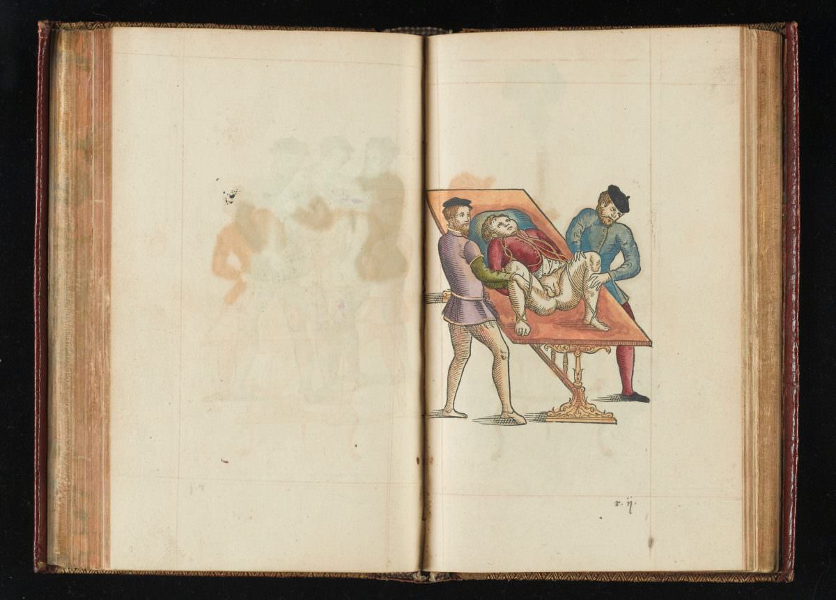
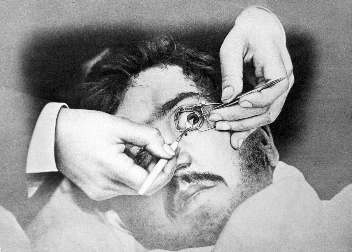
Imagine a world before antibiotics, anesthesia, and sterile operating rooms.  A world where surgery was a last resort, often leading to more suffering or even death.  This was the reality of the 19th century, a time of both medical advancement and brutal realities.  Thankfully, we have moved beyond those times, but the illustrations from that era provide a fascinating glimpse into the history of surgery.

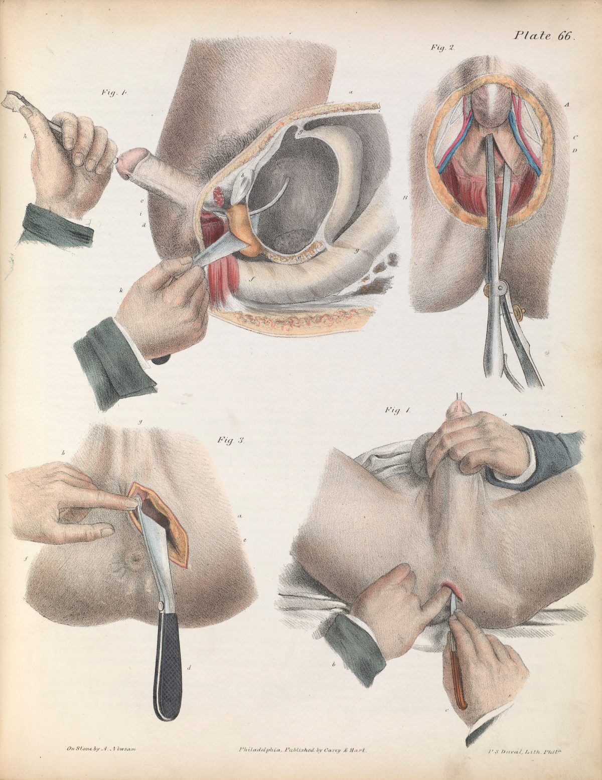
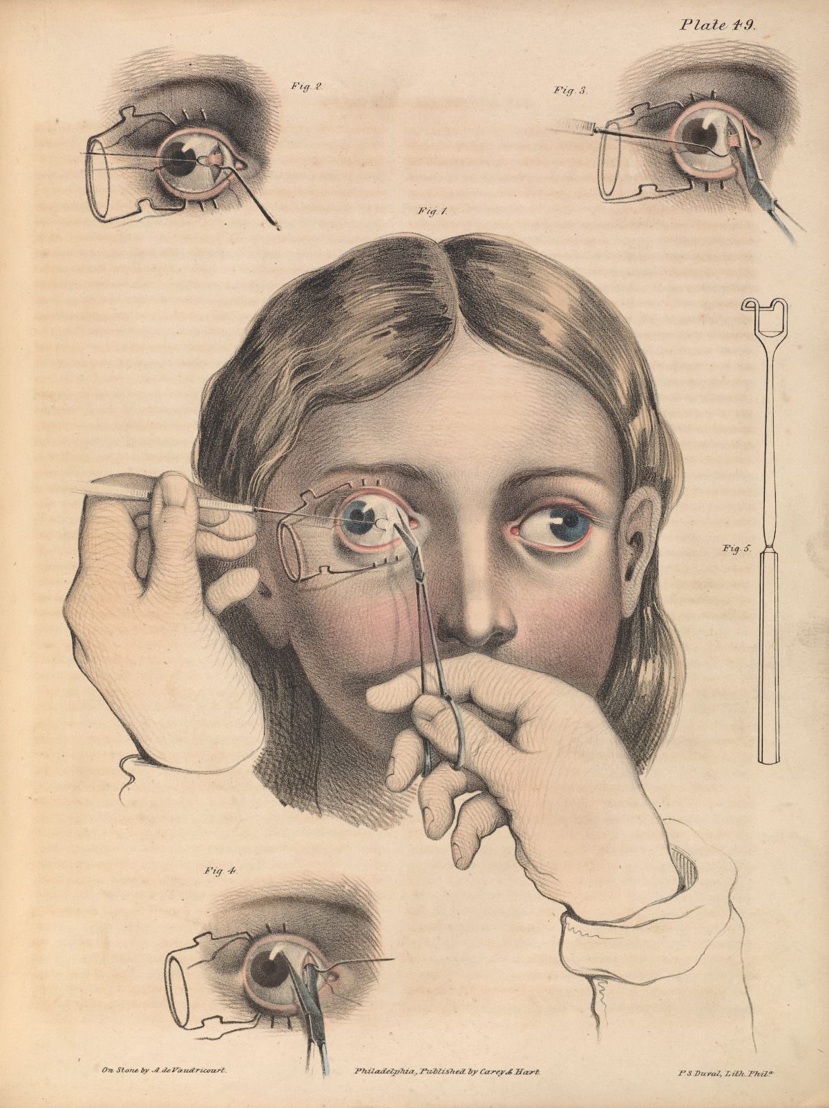
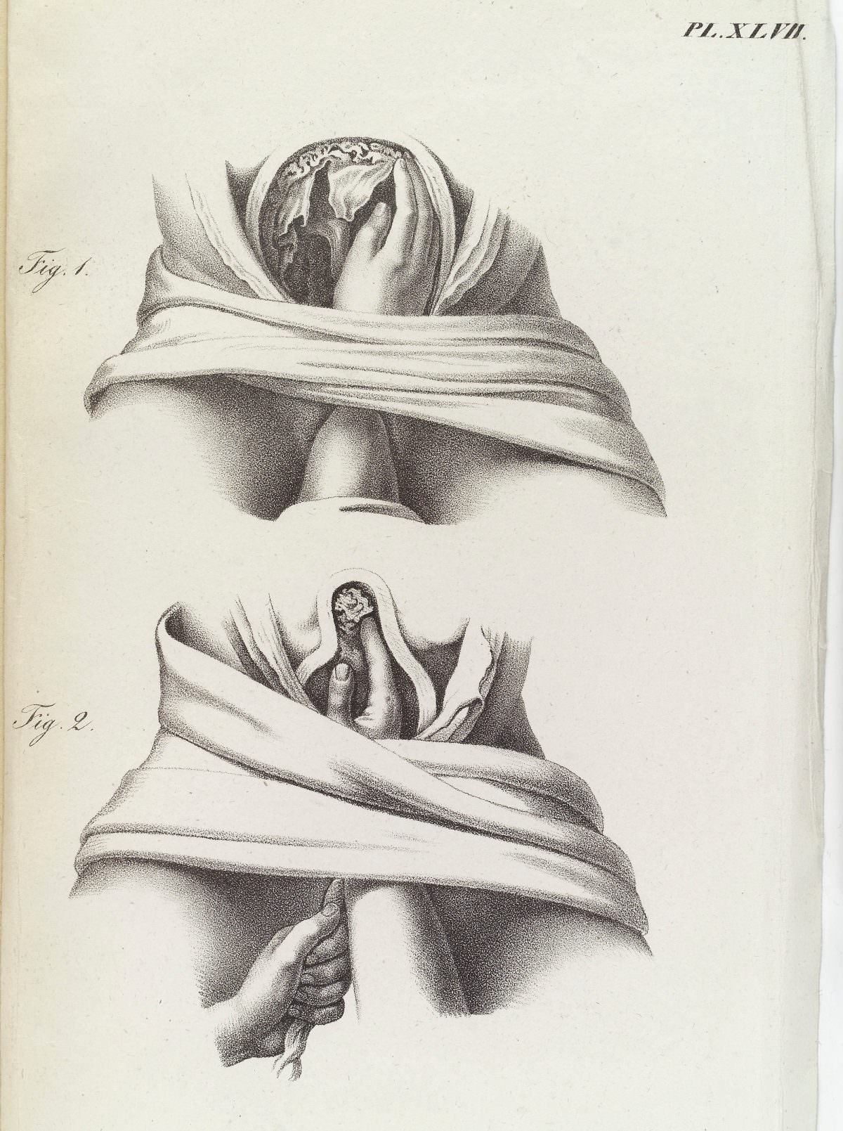
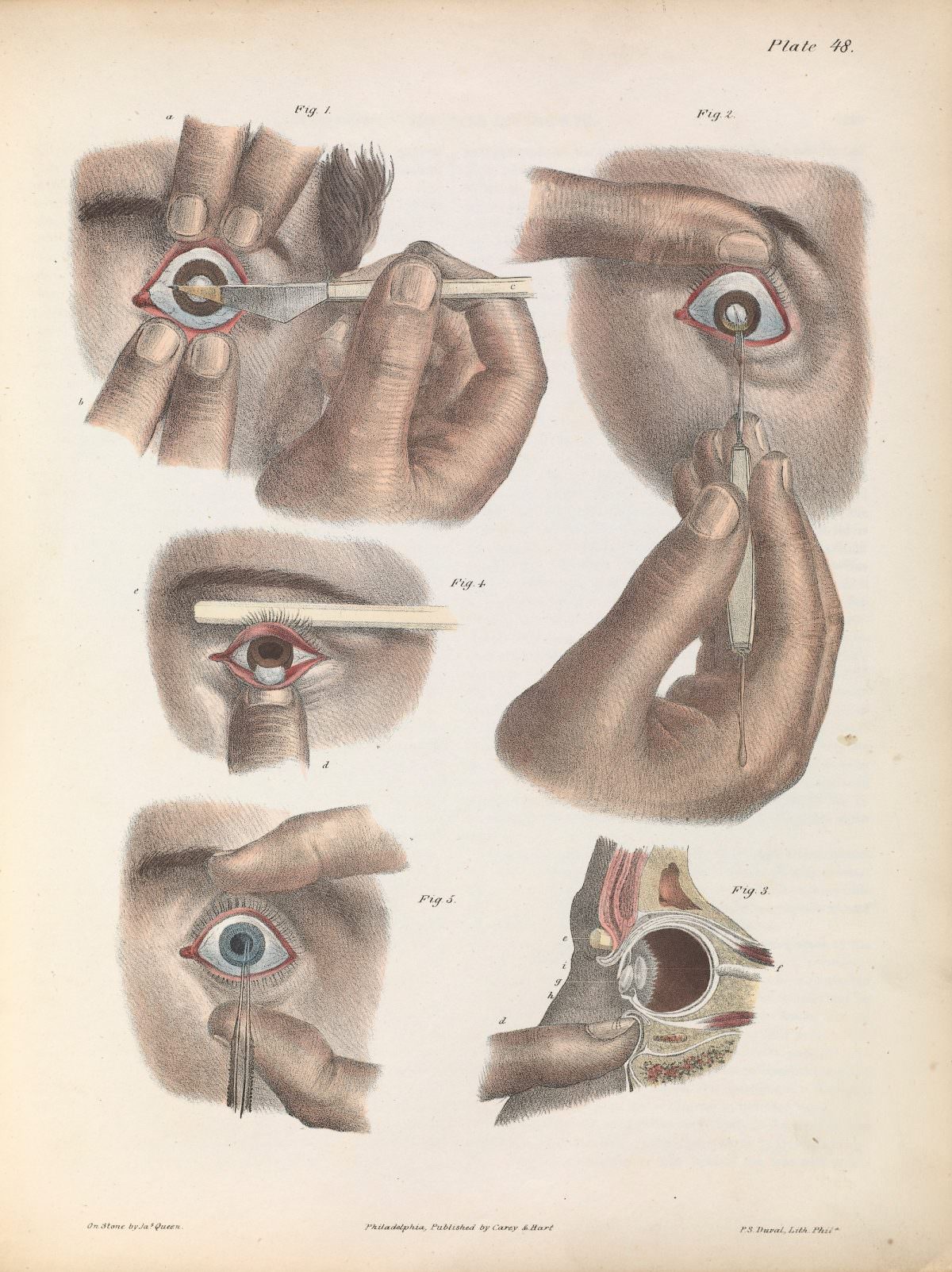
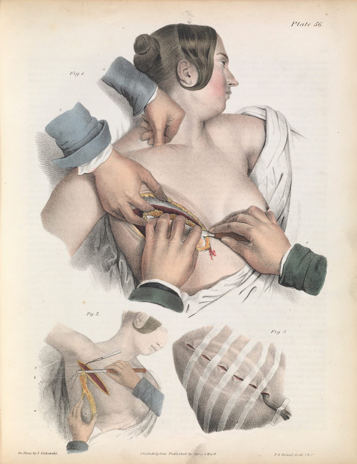
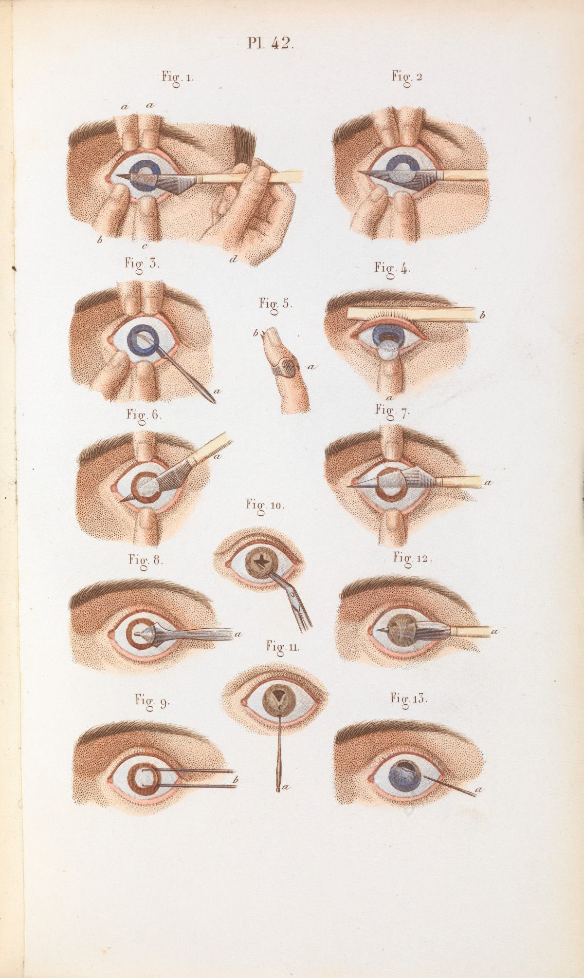
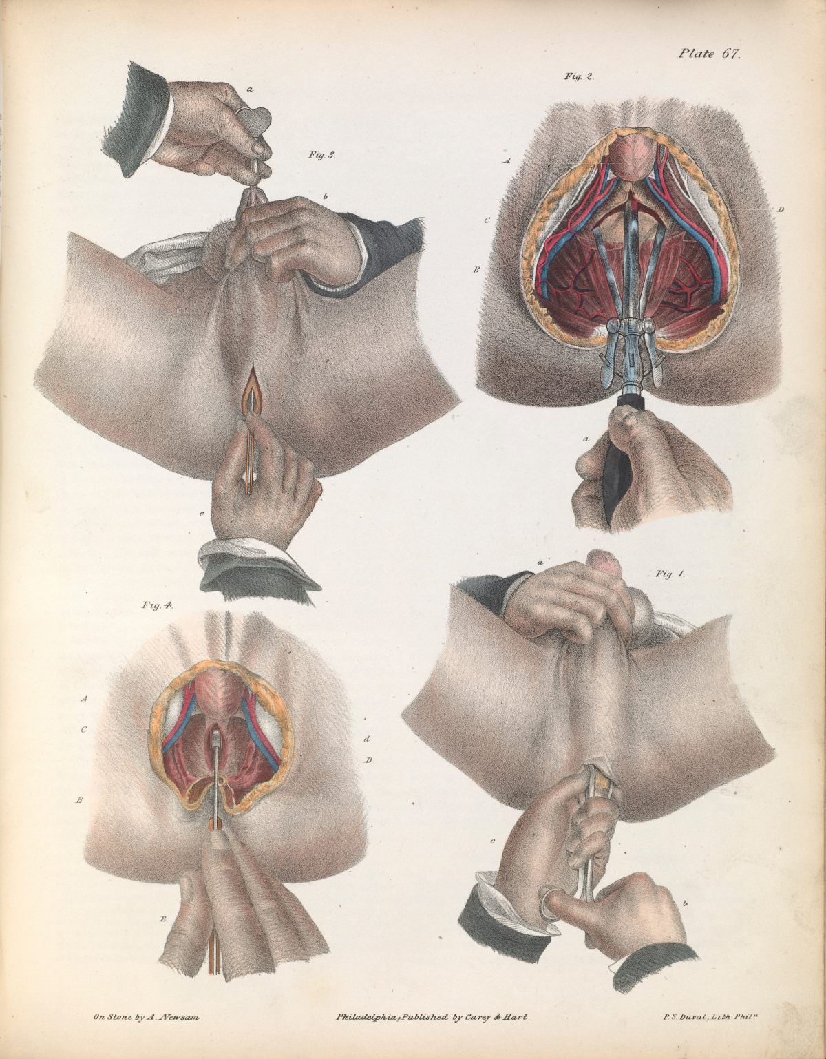
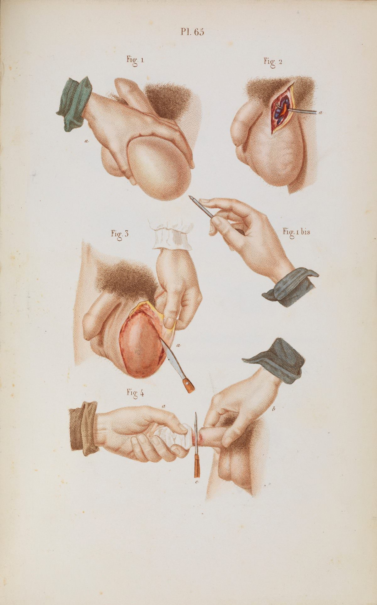
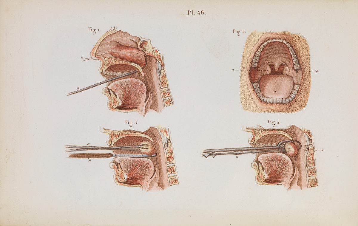
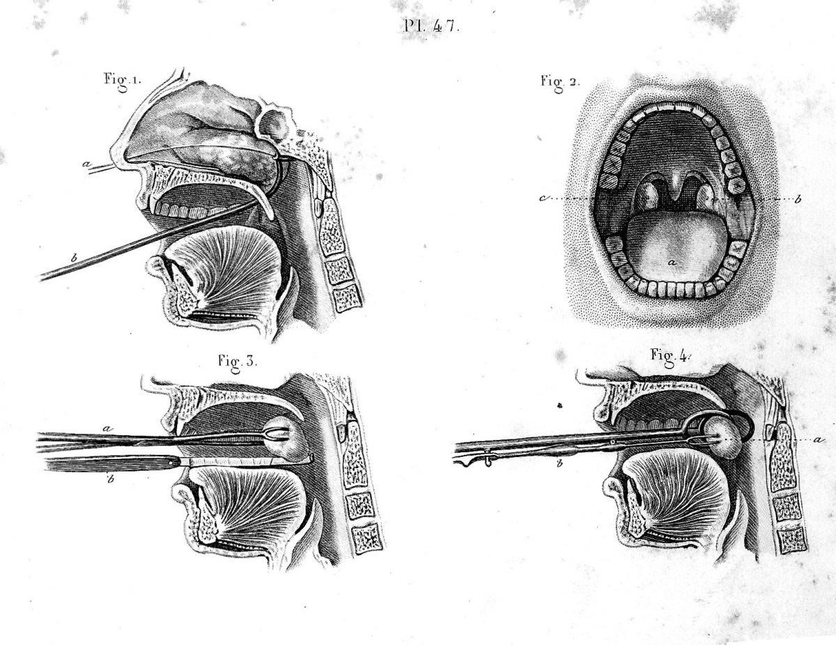
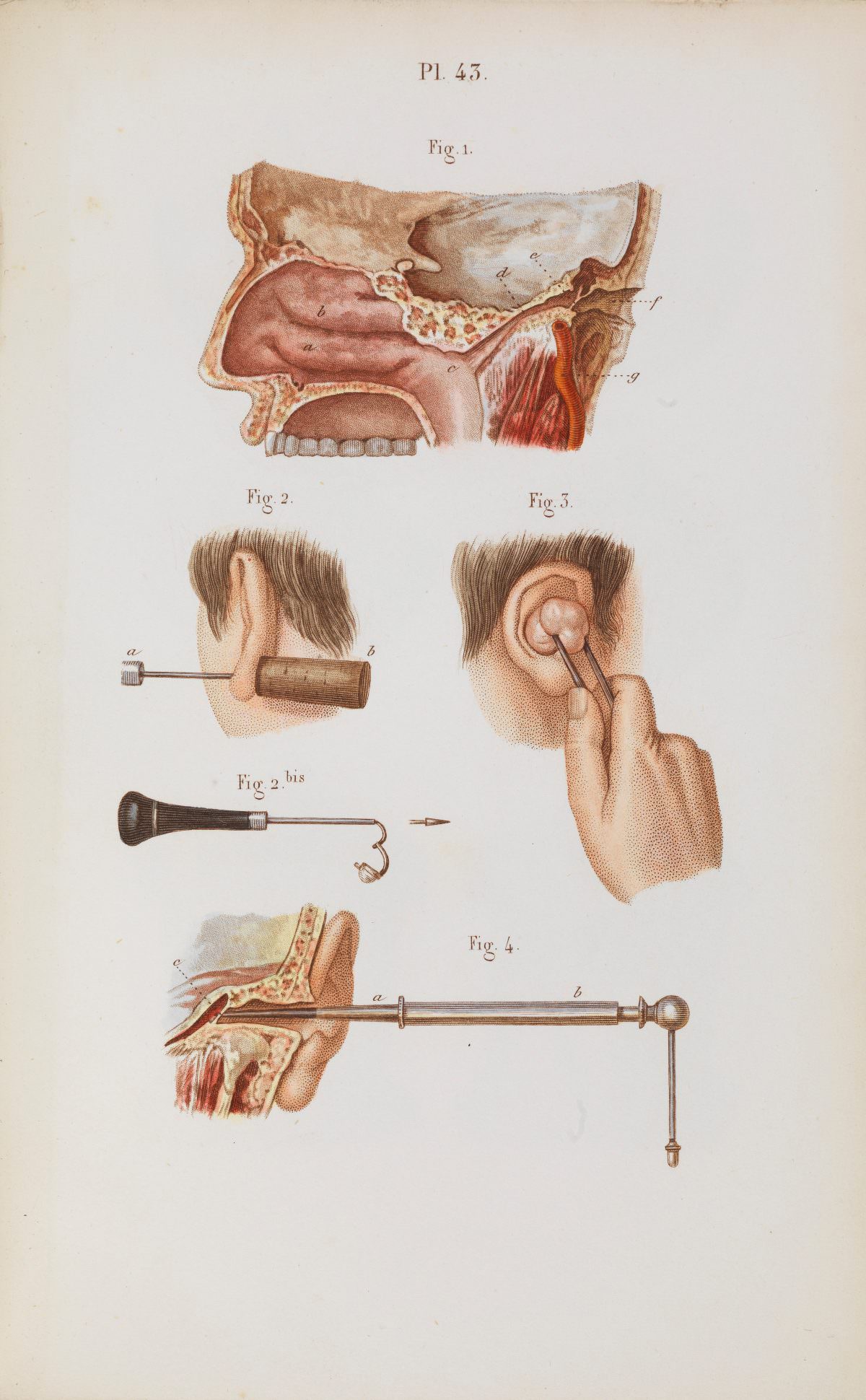
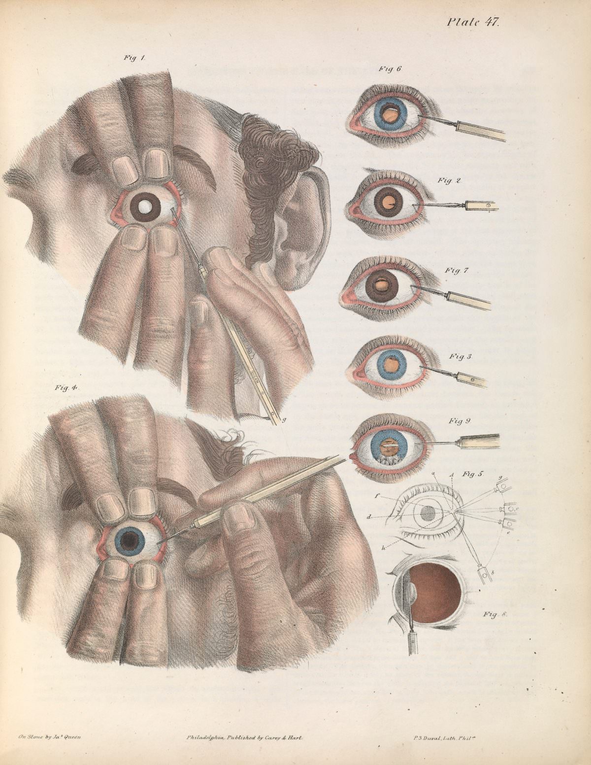
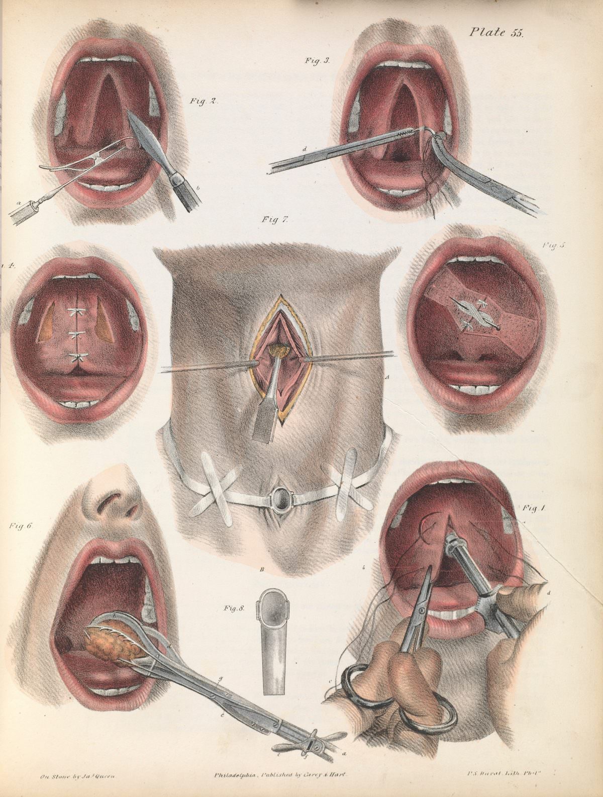
One such collection of illustrations comes from American surgeon Joseph Pancoast’s 1844 book, “A Treatise on Operative Surgery.”  The book, with its detailed descriptions and vivid illustrations, aimed to educate other surgeons on the latest techniques.  These images, while captivating, can be quite unsettling to the modern eye.

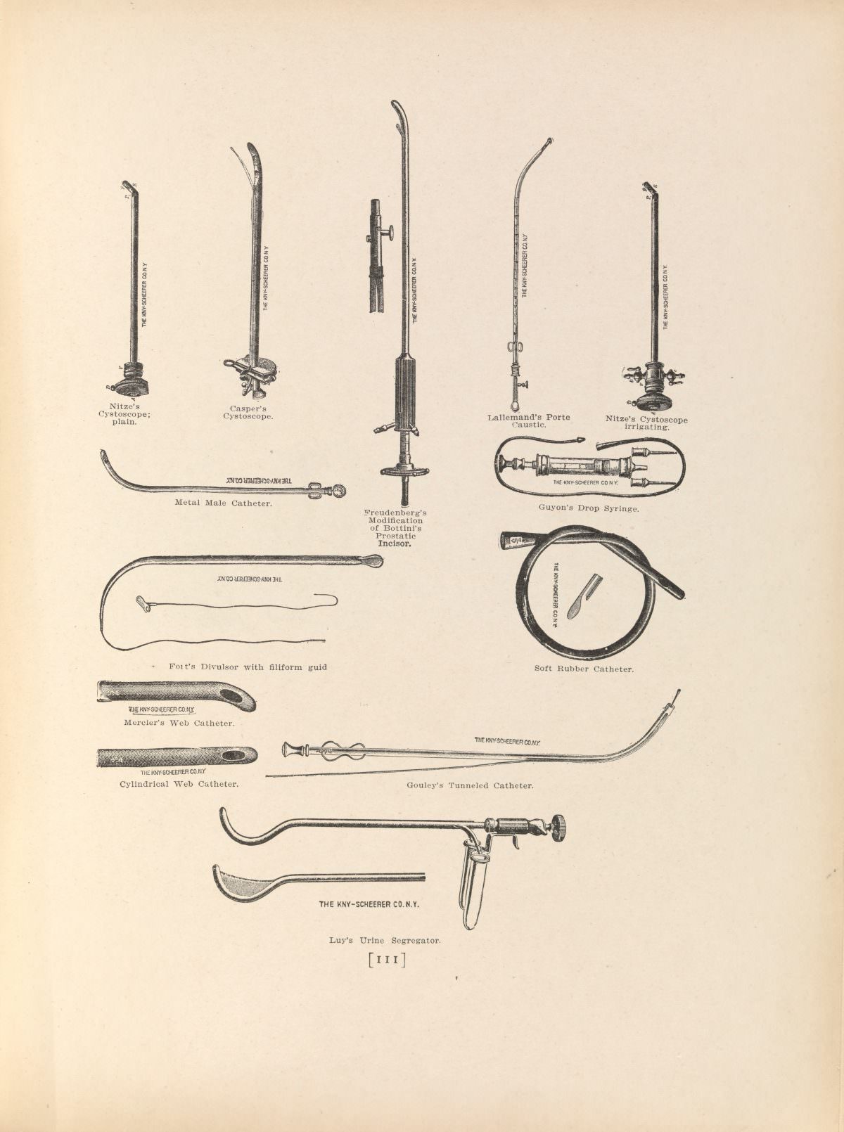
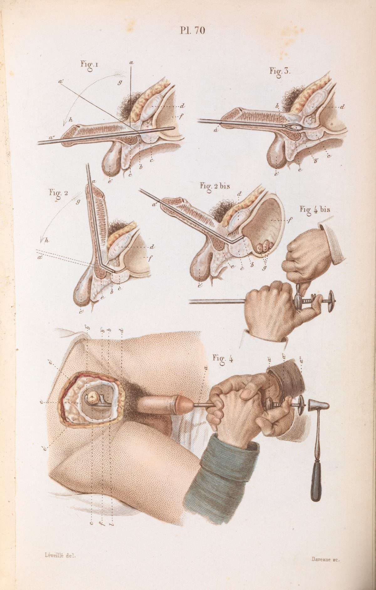
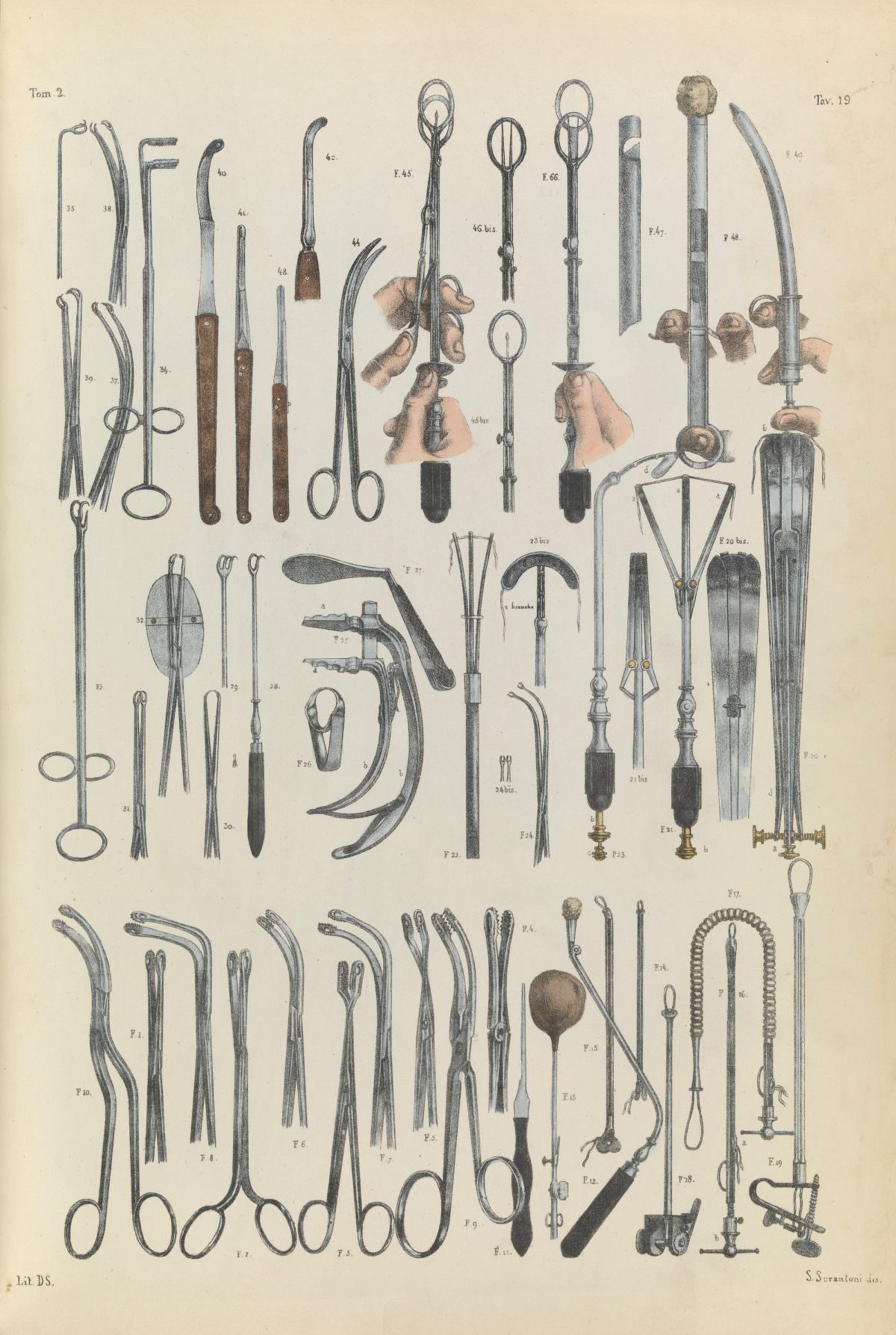
The illustrations showcase a variety of surgical instruments that, compared to today’s sleek and precise tools, appear rather crude and intimidating.  Scalpels, saws, forceps, and needles are depicted in detail, reminding us of the rawness and physicality of 19th-century surgery.  Many of these instruments were designed for amputations, a common procedure at the time due to war injuries and the lack of effective treatments for infections.

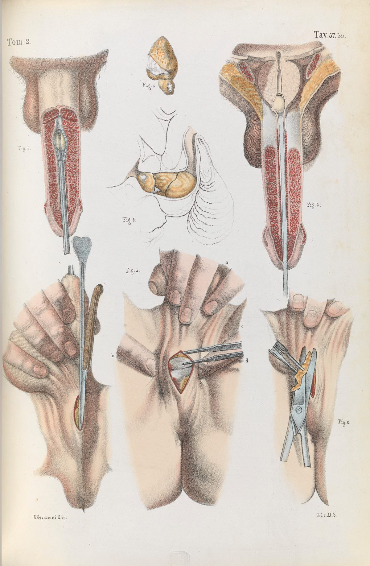
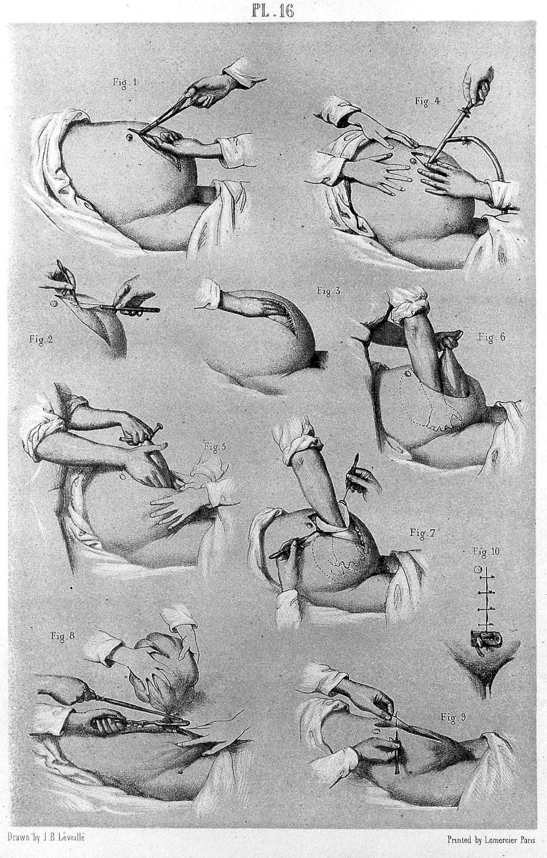
Pancoast’s illustrations go beyond just showcasing tools; they depict the human body and the surgical procedures themselves.  We see images of limbs being amputated, tumors being removed, and even a cleft palate surgery.  While the anatomical details are impressive, the exposed muscles, bones, and blood vessels are a stark reminder of the pain and risk involved in these procedures.

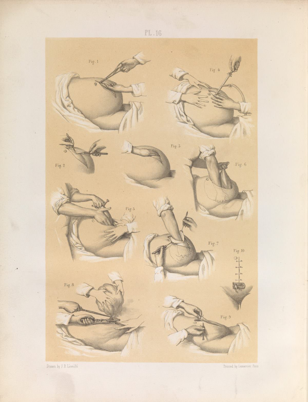
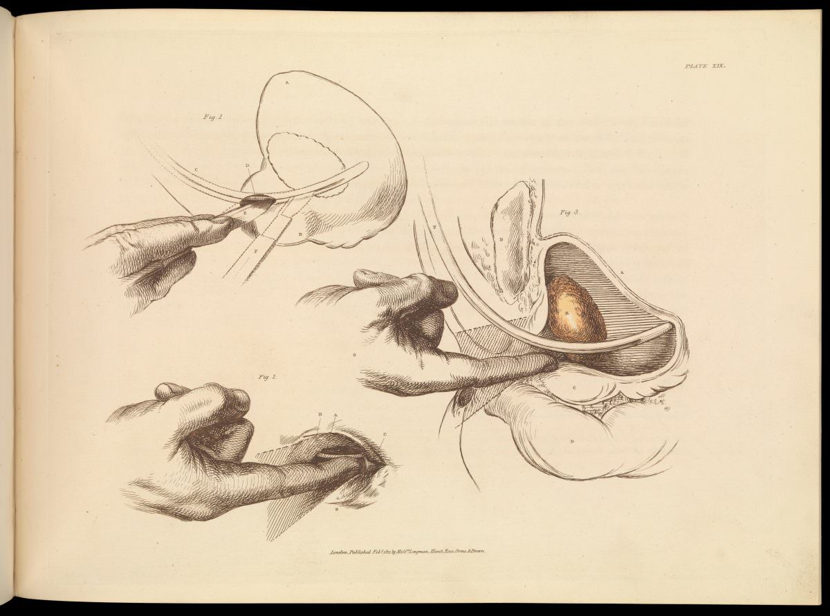
In 1848, French physiologist Claude Bernard published “Précis iconographique de médecine opératoire et d’anatomie chirurgicale” (Iconographic précis of operative medicine and surgical anatomy).  This work also included detailed illustrations of various surgeries, further adding to the visual record of 19th-century medical practices.

These illustrations, though sometimes unsettling, offer valuable insights into the history of medicine. They remind us of how far we’ve come in terms of surgical techniques, pain management, and overall patient care.  They also highlight the bravery of both patients and surgeons who faced the challenges of surgery in a time with limited resources and understanding of the human body.

#1 Plate LXVI. Surgical technique for lithotomy.

Plate LXVI. Surgical technique for lithotomy.

Leave a Reply

19th Century Illustrations for the Surgical Removal of Unwanted Parts of the Human Body

Your email address will not be published. Required fields are marked *

#2 Plate XLIX. Surgery to correct strabismus, involving the division of the internal rectus of the right eye. Strabismus is the misalignment of the eyes.

Plate XLIX. Surgery to correct strabismus, involving the division of the internal rectus of the right eye. Strabismus is the misalignment of the eyes.

Leave a Reply

19th Century Illustrations for the Surgical Removal of Unwanted Parts of the Human Body

Your email address will not be published. Required fields are marked *

#3 Plate XXXIV. Surgical instruments used for external urethrotomy in prostatectomy (removal of part of the prostate gland).

Plate XXXIV. Surgical instruments used for external urethrotomy in prostatectomy (removal of part of the prostate gland).

Leave a Reply

19th Century Illustrations for the Surgical Removal of Unwanted Parts of the Human Body

Your email address will not be published. Required fields are marked *

#4 Removing the placenta and umbilical cord after birth. Abbildungen aus dem Gesammtgebiete der theoretisch-praktischen Geburtshülfe, nebst beschreibender Erklärung derselben / Nach dem Französischen des Maygrier bearbeitet und mit Anmerkungen versehen von Eduard Casp. Jac. von Siebold – 1829

Removing the placenta and umbilical cord after birth. Abbildungen aus dem Gesammtgebiete der theoretisch-praktischen Geburtshülfe, nebst beschreibender Erklärung derselben / Nach dem Französischen des Maygrier bearbeitet und mit Anmerkungen versehen von Eduard Casp. Jac. von Siebold – 1829

Leave a Reply

19th Century Illustrations for the Surgical Removal of Unwanted Parts of the Human Body

Your email address will not be published. Required fields are marked *

#5 Plate XLVIII. Illustration of surgery on the eye for the removal of a cataract. Operation by extraction – inferior section of the cornea.

Plate XLVIII. Illustration of surgery on the eye for the removal of a cataract. Operation by extraction – inferior section of the cornea.

Leave a Reply

19th Century Illustrations for the Surgical Removal of Unwanted Parts of the Human Body

Your email address will not be published. Required fields are marked *

#6 Plate LVI. Surgery for the removal of the mammary gland. 19th Century. Iconografia d’anatomia chirurgica e di medicina operatoria

Plate LVI. Surgery for the removal of the mammary gland. 19th Century. Iconografia d’anatomia chirurgica e di medicina operatoria

Leave a Reply

19th Century Illustrations for the Surgical Removal of Unwanted Parts of the Human Body

Your email address will not be published. Required fields are marked *

#7 Plate 42, Techniques for the removal of cataracts.

Plate 42, Techniques for the removal of cataracts.

Leave a Reply

19th Century Illustrations for the Surgical Removal of Unwanted Parts of the Human Body

Your email address will not be published. Required fields are marked *

#8 Plate 70, Surgical techniques for lithotripsy (the removal of bladder and kidney stones). Précis iconographique de médecine opératoire et d’anatomie chirurgicale by Claude Bernard (1848).

Plate 70, Surgical techniques for lithotripsy (the removal of bladder and kidney stones). Précis iconographique de médecine opératoire et d’anatomie chirurgicale by Claude Bernard (1848).

Leave a Reply

19th Century Illustrations for the Surgical Removal of Unwanted Parts of the Human Body

Your email address will not be published. Required fields are marked *

#9 Plate LXVII. Surgical technique for lithotomy (the removal of a bladder stone). Bilateral and vesico-rectal operation.

Plate LXVII. Surgical technique for lithotomy (the removal of a bladder stone). Bilateral and vesico-rectal operation.

Leave a Reply

19th Century Illustrations for the Surgical Removal of Unwanted Parts of the Human Body

Your email address will not be published. Required fields are marked *

#10 Fig. 1. Removal with the forceps by torsion and traction. Fig. 2, 3. Removal by ligature. Fig. 4,5. Simple hare-lip. Fig. 6, 7, 8. ‘Double hare-lip’ and ‘complicated hare-lip’.

Fig. 1. Removal with the forceps by torsion and traction. Fig. 2, 3. Removal by ligature. Fig. 4,5. Simple hare-lip. Fig. 6, 7, 8. ‘Double hare-lip’ and ‘complicated hare-lip’.

Leave a Reply

19th Century Illustrations for the Surgical Removal of Unwanted Parts of the Human Body

Your email address will not be published. Required fields are marked *

#11 Plate 65, Surgical removal of tumours from the scrotum.

Plate 65, Surgical removal of tumours from the scrotum.

Leave a Reply

19th Century Illustrations for the Surgical Removal of Unwanted Parts of the Human Body

Your email address will not be published. Required fields are marked *

#12 Plate 46, Illustration of the removal of nasal polyps and tonsillectomy.

Plate 46, Illustration of the removal of nasal polyps and tonsillectomy.

Leave a Reply

19th Century Illustrations for the Surgical Removal of Unwanted Parts of the Human Body

Your email address will not be published. Required fields are marked *

#13 Plate 16. Various operative stages of the removal of a tumour from the uterus.

Plate 16. Various operative stages of the removal of a tumour from the uterus.

Leave a Reply

19th Century Illustrations for the Surgical Removal of Unwanted Parts of the Human Body

Your email address will not be published. Required fields are marked *

#14 Tomo II, Tavola 19. Surgical instruments used on the tonsils and nasal cavity for the removal of polyps.

Tomo II, Tavola 19. Surgical instruments used on the tonsils and nasal cavity for the removal of polyps.

Leave a Reply

19th Century Illustrations for the Surgical Removal of Unwanted Parts of the Human Body

Your email address will not be published. Required fields are marked *

#15 Tomo II, Tavola 57 bis. Procedure for the removal of stones from the prostate and urethra.

Tomo II, Tavola 57 bis. Procedure for the removal of stones from the prostate and urethra.

Leave a Reply

19th Century Illustrations for the Surgical Removal of Unwanted Parts of the Human Body

Your email address will not be published. Required fields are marked *

#16 Removal of tumours of uterus

Removal of tumours of uterus

Leave a Reply

19th Century Illustrations for the Surgical Removal of Unwanted Parts of the Human Body

Your email address will not be published. Required fields are marked *

#17 Hand painted wood engraving showing the necessary position for the patient for removal of bladder stones.

Hand painted wood engraving showing the necessary position for the patient for removal of bladder stones.

Leave a Reply

19th Century Illustrations for the Surgical Removal of Unwanted Parts of the Human Body

Your email address will not be published. Required fields are marked *

#18

19th Century Illustrations for the Surgical Removal of Unwanted Parts of the Human Body

Leave a Reply

19th Century Illustrations for the Surgical Removal of Unwanted Parts of the Human Body

Your email address will not be published. Required fields are marked *

#19 Surgery on the eye for the removal of a cataract.

Surgery on the eye for the removal of a cataract.

Leave a Reply

19th Century Illustrations for the Surgical Removal of Unwanted Parts of the Human Body

Your email address will not be published. Required fields are marked *

#20 Plate XIX. Surgical removal of a stone from the bladder.

Plate XIX. Surgical removal of a stone from the bladder.

Leave a Reply

19th Century Illustrations for the Surgical Removal of Unwanted Parts of the Human Body

Your email address will not be published. Required fields are marked *

#21 Plate 43, Illustration and anatomy of ear surgery.

Plate 43, Illustration and anatomy of ear surgery.

Leave a Reply

19th Century Illustrations for the Surgical Removal of Unwanted Parts of the Human Body

Your email address will not be published. Required fields are marked *

#22 Removing cataracts

Removing cataracts

Leave a Reply

19th Century Illustrations for the Surgical Removal of Unwanted Parts of the Human Body

Your email address will not be published. Required fields are marked *

#23 Plate 55, J. Pancoast, A treatise on operative surgery, 1846.

Plate 55, J. Pancoast, A treatise on operative surgery, 1846.

Leave a Reply

19th Century Illustrations for the Surgical Removal of Unwanted Parts of the Human Body

Your email address will not be published. Required fields are marked *

Avatar of Megan Di

Written by Megan Di

Leave a Reply

Your email address will not be published. Required fields are marked *