
Modern surgery lets doctors transplant organs while a patient sleeps through the entire procedure. That reality would have seemed impossible to people living just a century or two ago. Before anesthesia was reliable, before germ theory was understood, and before medical equipment became precise, patients faced procedures that were often as dangerous as the conditions they were meant to fix. Many diseases had no real treatment at all, leaving patients to suffer until they died. The photos below document real instruments, real procedures, and real hospital conditions from the past — and they are not easy to look at.
One of the most extreme examples of medical desperation in history took place at a remote Soviet Antarctic research station. In 1961, the station’s only physician discovered he was suffering from acute appendicitis — a life-threatening condition requiring immediate surgery. With no other surgeon available, he operated on himself.
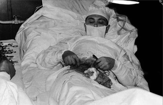
Dr. Leonid Rogozov made a 12-centimeter incision into his own abdomen, located the infected appendix, and removed it. Five days later he was recovering well. Seven days after the operation, he removed his own sutures. His case remains one of the most remarkable acts of self-surgery ever recorded.
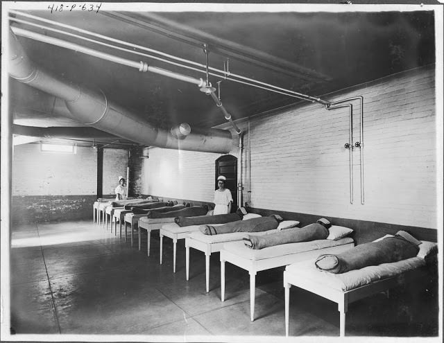
Mental institutions throughout the 19th and early 20th centuries had few effective treatments for psychiatric conditions. Restraint was one of the most common responses to patients who showed agitation or distress.
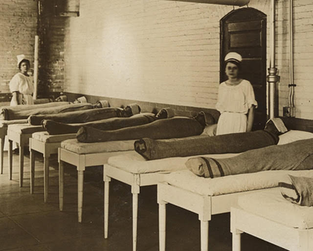
Wet blankets were wrapped tightly around patients to physically immobilize them. The practice was considered a form of hydrotherapy. Whether it calmed patients or simply trapped them, it was widely used across institutions in Europe and North America.
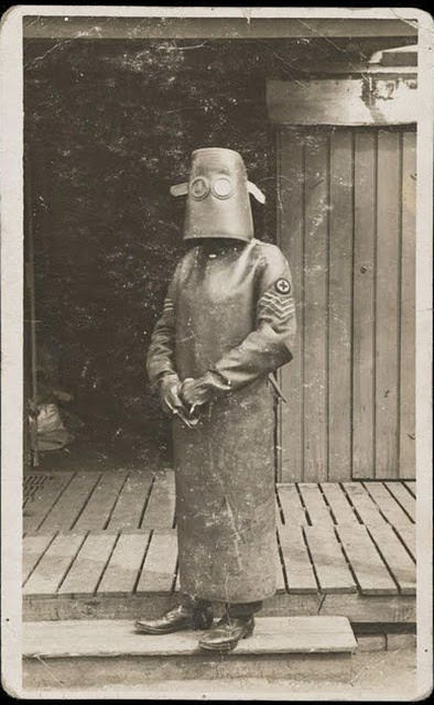
Early radiology workers faced serious dangers because the harm caused by X-ray radiation was not yet fully understood. Nurses and technicians wore bulky lead-lined gear to shield themselves, though the protection it offered was often incomplete. Dr. Maxime Menard, who ran a radiology department at a Paris hospital around the same era, later lost a finger to side effects from operating his X-ray machine.
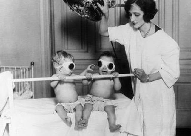
Rickets, caused by a vitamin D deficiency, was a serious problem for children in the early 20th century — especially in urban orphanages where access to sunlight and nutritious food was limited. In 1925, staff at the Chicago Orphan Asylum exposed infants to artificial ultraviolet light as a way to compensate for sunlight lost during winter months. The method reflected a growing but still rough understanding of how sunlight affected bone development.
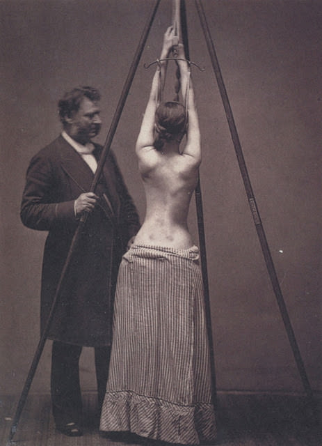
Scoliosis — an abnormal curvature of the spine — was a condition that baffled physicians for generations. Lewis Sayre was among the first American surgeons to tackle it systematically. His methods involved manual assessment and mechanical correction. Others devised more extreme approaches.
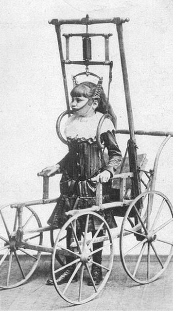
Dr. Clark’s Spinal Apparatus, introduced in 1878, was a mechanical device designed to pull the spine into alignment. It was widely considered one of the most impractical scoliosis treatments ever produced. Patients were subjected to significant discomfort with little measurable benefit.
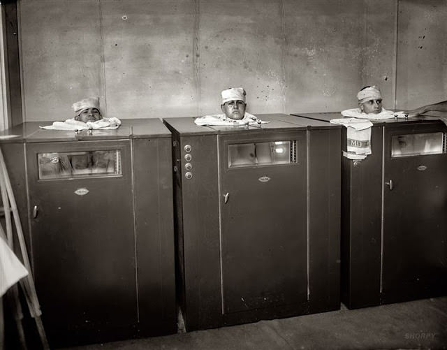
By 1920, Walter Reed Army Hospital was using mechanical devices for physiotherapy rather than direct hands-on treatment. Soldiers and patients recovering from injuries were connected to machines designed to move or stimulate muscles. The approach looked clinical but marked a genuine step toward structured physical rehabilitation.
Before anesthesia became standard, surgery was performed on fully conscious patients. Speed was considered the greatest skill a surgeon could have — the faster the procedure, the less the patient suffered.
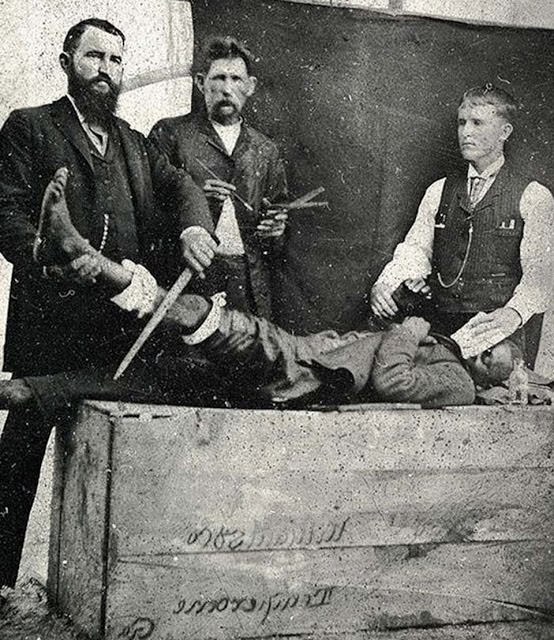
This 1855 photograph shows a young patient undergoing leg surgery without anesthesia. Patients were often held down by assistants or strapped to a table. Screaming during the procedure was not unusual. Early ether and chloroform anesthesia existed by this point but were not yet universally adopted.
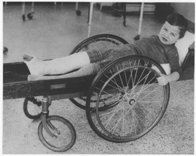
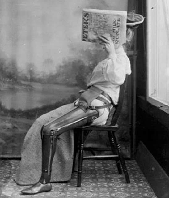
Artificial limbs in the 1890s were functional at best and disfiguring at worst. Many amputees, especially women, faced intense social stigma. This photograph shows a woman who chose to hide her face rather than be identified with her prosthetic leg — a reflection of how disability was viewed at the time, not just how it was treated.
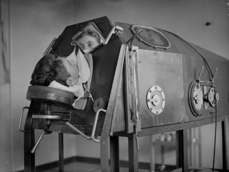
Polio was one of the most feared diseases of the 20th century. It attacked the nervous system and could leave patients unable to breathe on their own. The iron lung — a large metal chamber that used air pressure changes to force the lungs to expand and contract — kept patients alive when their respiratory muscles failed.
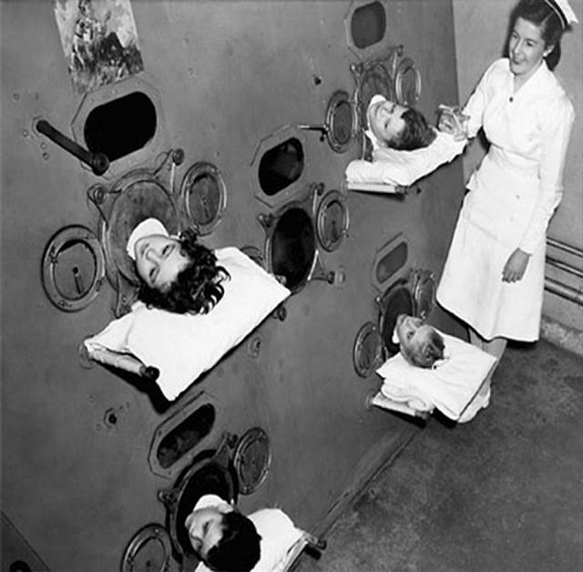
During major polio outbreaks, hospitals ran out of single-person iron lungs. Engineers responded by building multi-patient versions that could hold several people at once. Patients lay with their bodies sealed inside the machine and only their heads exposed, often for months or years at a time.
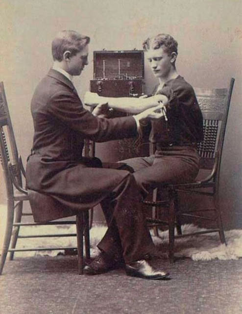
Neurological examinations in the 1880s relied almost entirely on observation and physical stimulus. Physicians tested reflexes, sensory responses, and muscular reactions by hand, with no imaging technology to confirm their findings. Diagnosis depended heavily on the individual doctor’s experience and judgment.
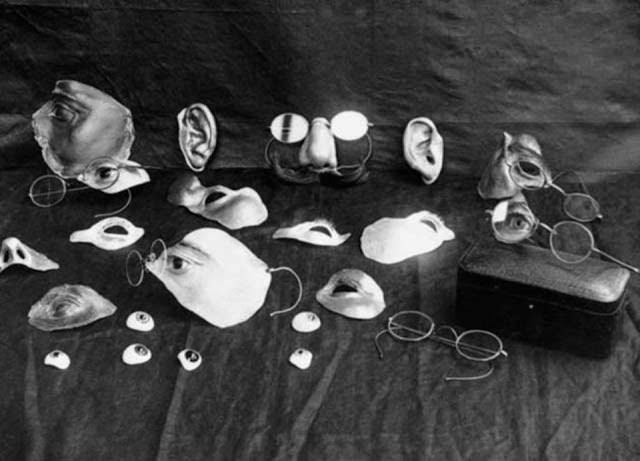
Before reconstructive plastic surgery became viable, soldiers and accident victims who suffered severe facial injuries had few options. Craftsmen created custom metal masks painted to match skin tones, designed to cover missing noses, cheeks, or eye sockets. These prosthetics allowed patients to move through public life without drawing immediate attention to their injuries.
Phrenology — the belief that the shape of a person’s skull revealed their mental abilities and character — was taken seriously by many in the early 20th century. Inventors even built machines to automate the process.
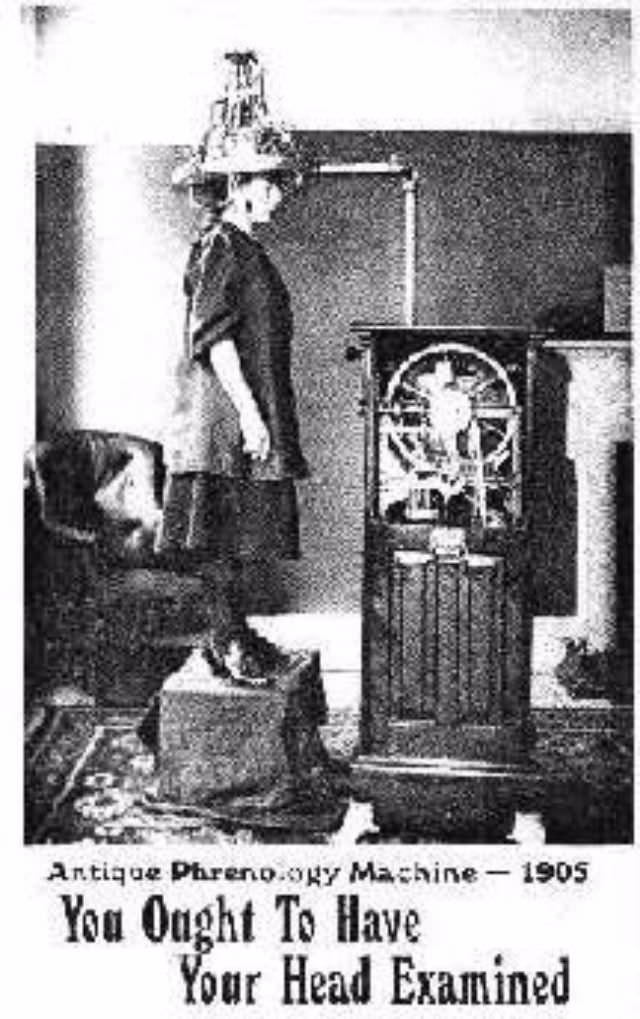
The Psychograph contained 1,954 parts housed in a walnut cabinet with a motor-driven belt. A headpiece with 32 probes pressed against the skull, sending low-voltage signals to the machine. The machine then stamped out printed statements rating 32 supposed mental faculties on a scale from “deficient” to “very superior.” Subjects sat in a connected chair while the operator pulled a lever to activate the whole system. It was presented as science.
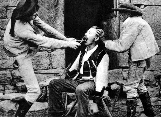
Early dentists operated under the belief that blood needed to flow before healing could begin. Extractions were performed with forceps and a penknife, with no anesthesia. The dentist’s apron was typically already bloodstained before a new patient sat down. Pain was simply part of the process.
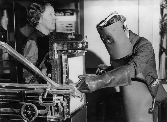
By 1934, X-ray technology had improved enough that the heavy protective gear worn by earlier radiologists was becoming obsolete. At a London radiological exhibition that year, a technician demonstrated the new equipment while still wearing the older style of protection — a visual reminder of how quickly the field was evolving.
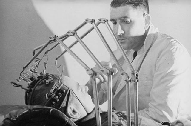
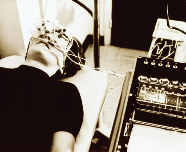
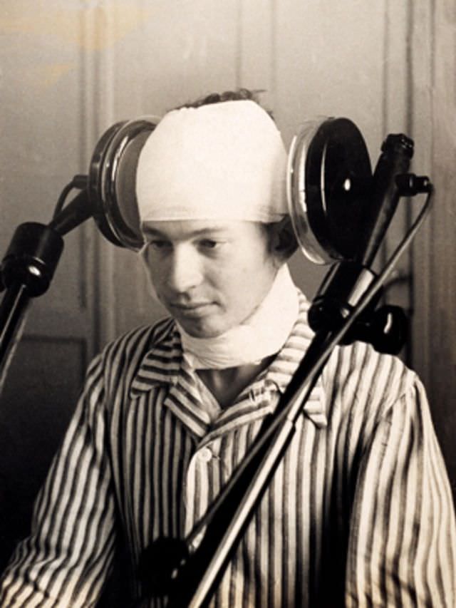
Early electroencephalography (EEG) required patients to wear a headpiece covered in electrodes connected to bulky recording equipment. The machines translated electrical activity in the brain into lines on paper. The setup was cumbersome and the readings were difficult to interpret, but it was the first time doctors could observe brain activity without opening the skull.
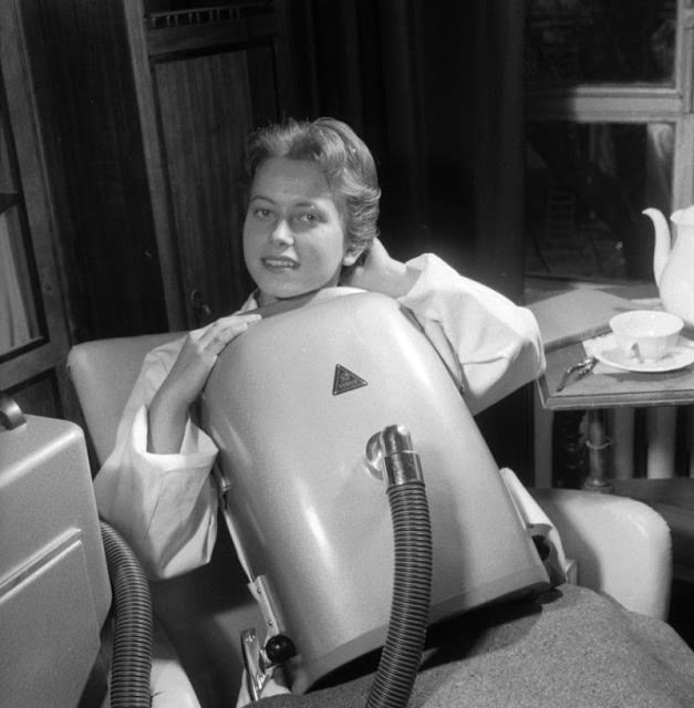
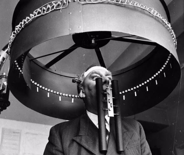
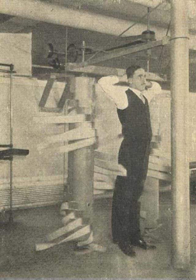
Aviation medicine pushed researchers to study how the human body responded to extreme conditions. At the RAF Institute of Aviation Medicine in Farnborough, Dr. G. H. Byford wore a contact lens with a miniature lamp attached directly to it while standing beneath an optokinetic drum — a rotating device used to trigger eye movement reflexes. The goal was to understand how visual illusions affected pilots.
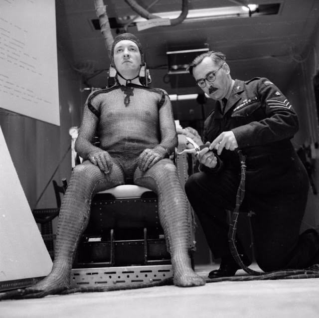
As the space race accelerated in 1960, researchers needed to understand how the human body coped with extreme speeds and the vacuum of space. This wire suit was designed to monitor body temperatures across different regions simultaneously, giving scientists data on how heat moved through the body under those conditions.
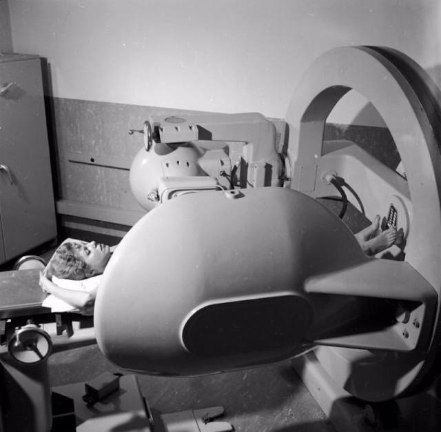
Cobalt-60 radiation therapy became one of the primary cancer treatments in the mid-20th century. The machine rotated around the patient’s body, directing beams of radiation at tumors from multiple angles to maximize damage to the cancer while reducing harm to surrounding tissue. It was a major advancement over earlier, static radiation devices.
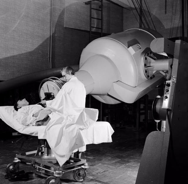
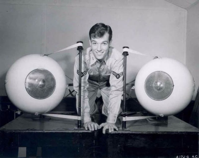
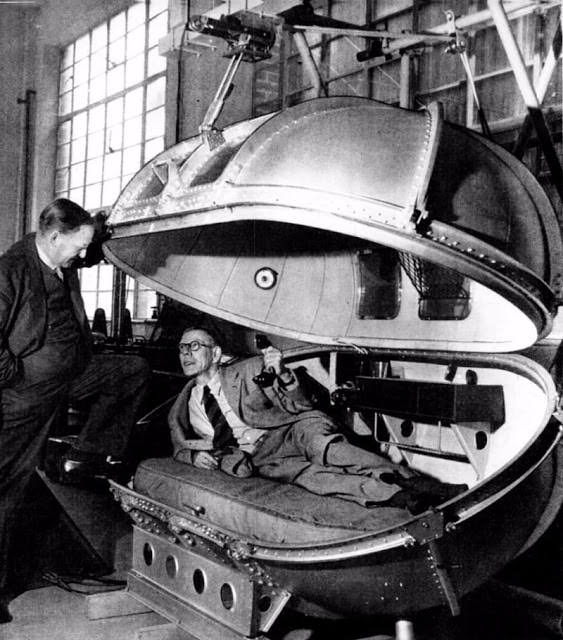
High-altitude flight posed serious health risks — including hypoxia and decompression sickness — that were not fully manageable with standard aircraft cabins during World War II. Winston Churchill’s personal pressurized chamber was built so he could safely travel at altitude without risking his health during critical wartime flights.
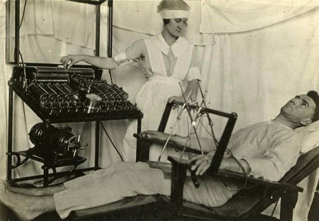
The Bergonic chair delivered electric current through a patient’s body as a treatment for psychological conditions. Patients sat in the chair and electrodes made contact with their limbs, sending mild to moderate electrical shocks through the nervous system. It was used specifically in cases described as “psycho-neurotic” — a broad and loosely defined category in early 20th century psychiatry.
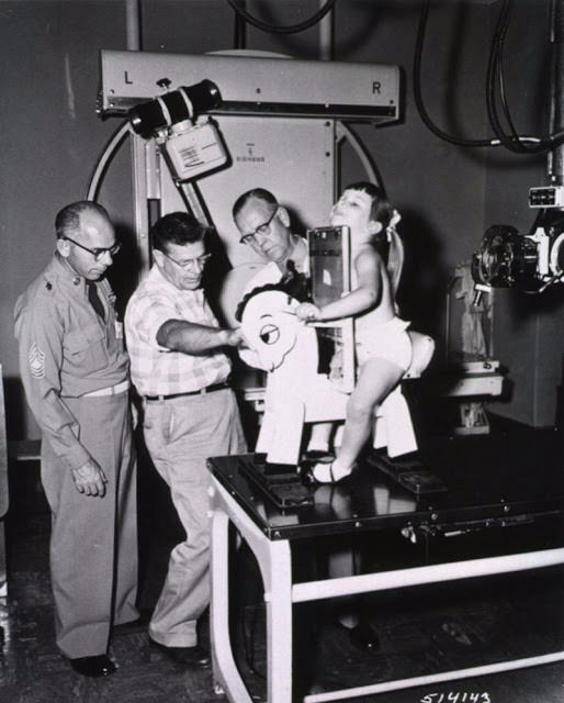
Getting young children to sit still long enough for a chest X-ray was a persistent challenge for pediatric radiologists. The Roentgen steed solved the problem by seating children on a horse-shaped frame that kept them upright and correctly positioned without requiring cooperation from the child.
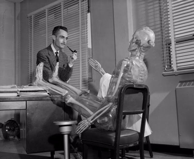
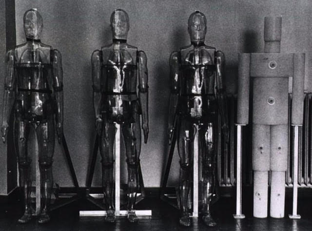
During the Cold War, researchers at Los Alamos needed to study how radiation penetrated and dispersed through the human body without exposing actual people. Plastic Man — a life-sized humanoid phantom filled with materials that mimicked human tissue — allowed scientists to measure radiation doses in ways that would have been impossible and unethical to perform on living subjects.
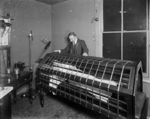
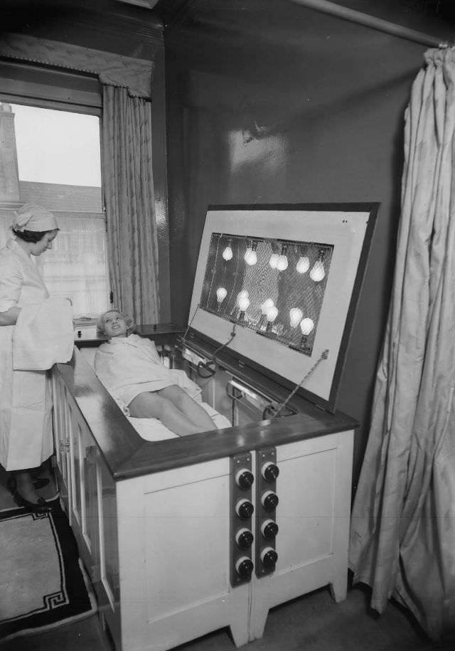
The Electric Bath was a treatment offered at light care institutes in the early 1900s. Patients sat inside a cabinet lined with electric light bulbs that generated heat and light across the body. It was marketed for a wide range of conditions and is considered a precursor to the modern sunbed, though its medical claims had no solid scientific basis.
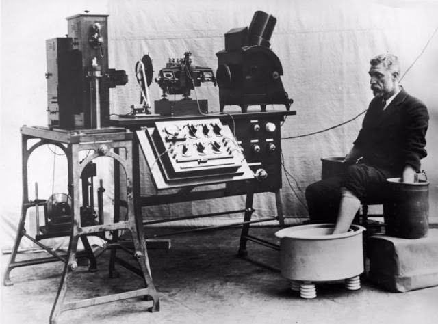
The electrocardiograph was one of the most genuinely useful diagnostic tools to emerge from early electrical medicine. Cambridge Scientific Instruments introduced one of the first commercial models, which measured the electrical activity of the heart and printed the results as a waveform. It formed the foundation of cardiac diagnostics that is still used today.
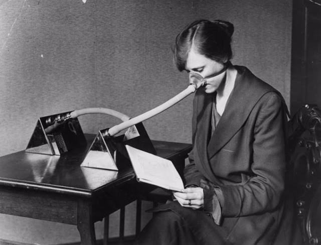
The influenza pandemic that followed World War I killed between 50 and 100 million people worldwide between 1918 and 1920. In the absence of effective vaccines or antiviral drugs, cloth face masks were distributed as a public health measure. They provided minimal protection, but they were among the few tools available.
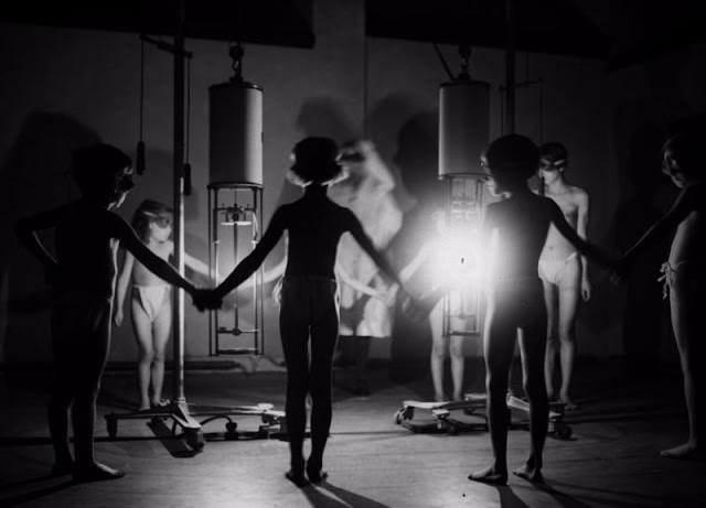
Ultraviolet light therapy was used in the early 20th century to treat a range of conditions, including tuberculosis of the skin, rickets, and various infections. Children gathered around UV lamps at specialized institutes, where the treatment was administered under medical supervision. The approach had genuine clinical backing in some applications.
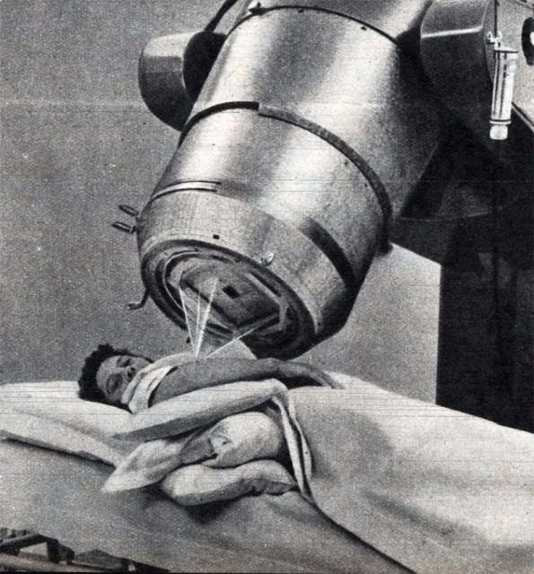
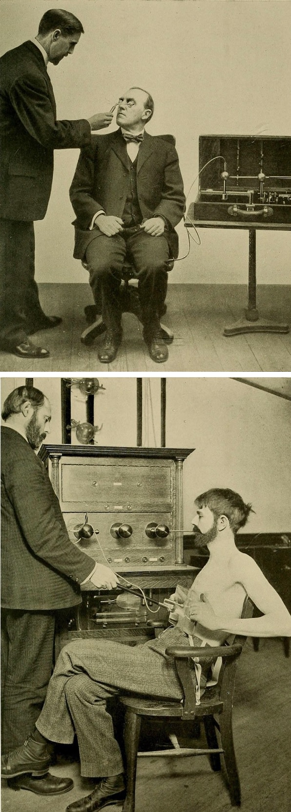
High-frequency electric current therapy was promoted in the early 20th century as a treatment for acne, insomnia, abnormal blood pressure, depression, hysteria, and lesions. The idea that electricity could heal the body stretched back to ancient Greeks using electric eels to numb pain, but it gained real traction in the 18th and 19th centuries through the work of Luigi Galvani and Guillaume Duchenne. By the 1900s, machines delivering these currents were being sold commercially to both physicians and patients.
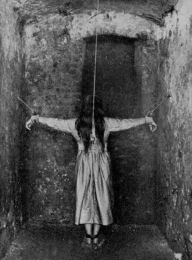
Psychiatric institutions used physical restraint as a routine management tool well into the 20th century. The devices varied in form, but the intent was consistently the same — to prevent patients from moving freely.
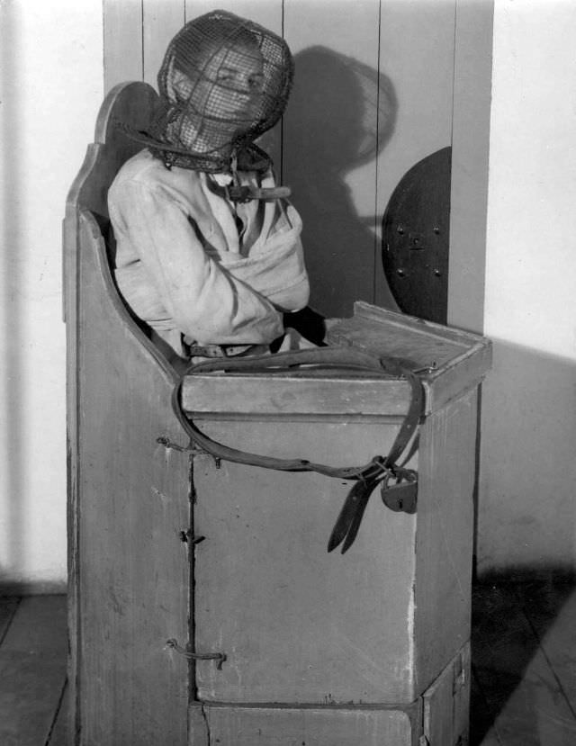
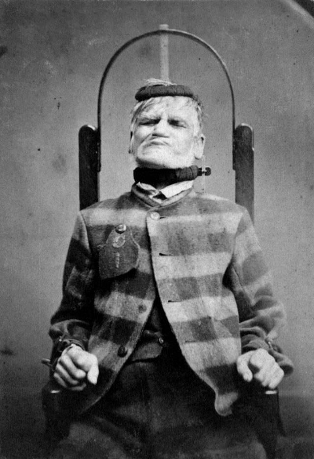
The restraining chair at the West Riding Lunatic Asylum in 1869 was designed to immobilize patients completely. Straps held the arms, legs, and torso in place. Patients could be kept in these chairs for hours at a time. The asylum’s records from this period show that restraint was used not only for violent patients but also for those who were simply uncooperative.
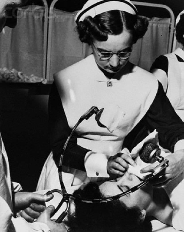
Electroconvulsive therapy (ECT) was introduced in the late 1930s and became widely used in psychiatric hospitals throughout the 1940s and 1950s. Electrodes were placed on the patient’s head and an electric current was passed through the brain, inducing a seizure. In early applications, patients were not given muscle relaxants, meaning the convulsions could be violent enough to cause broken bones.
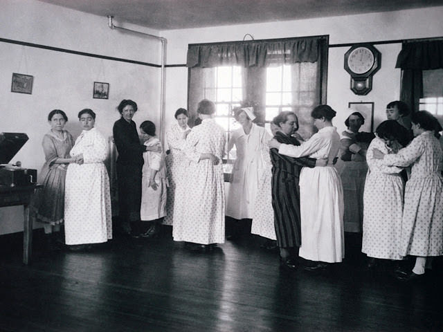
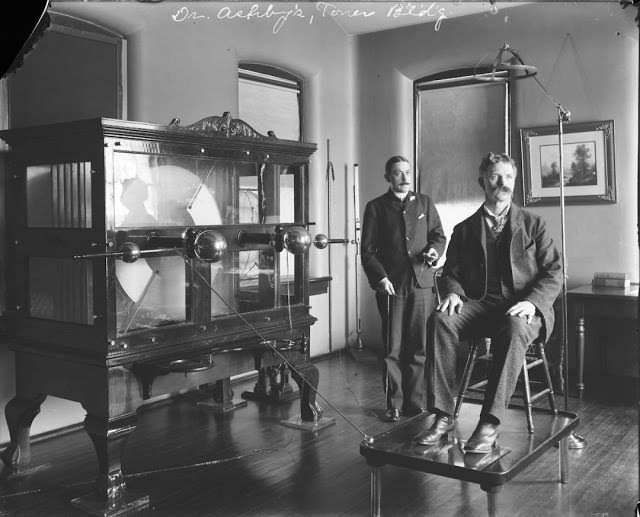
St. Elizabeths Hospital in Washington, D.C. was one of the first major psychiatric institutions in the United States. By 1923, its medical staff were using a large electro-shock machine — the size of a piece of heavy furniture — on patients. The doctors photographed here presented it as a medical advancement.
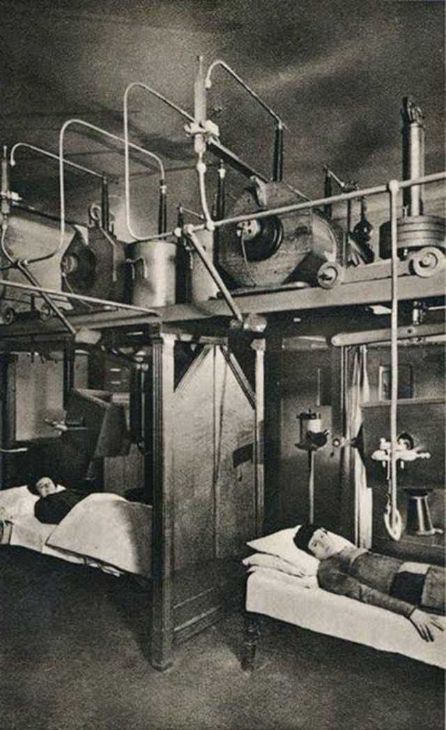
Radium was discovered in 1898 and was quickly marketed as a healing substance. By 1907, it was being used in a Serbian psychiatric hospital as a treatment for mental illness. The dangers of radium exposure — radiation sickness, cancer, and death — were not yet understood. Many patients and medical workers who were exposed to radium in this era suffered serious long-term harm.
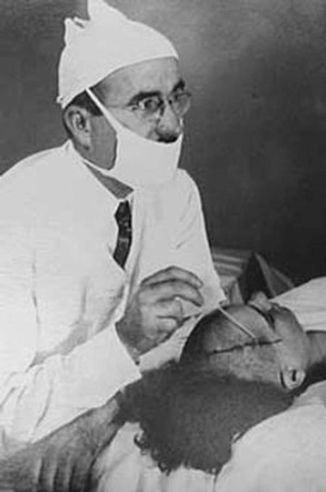
Hydrotherapy was used extensively in psychiatric hospitals as a calming treatment. Patients were submerged in tubs of water for anywhere from a few hours to an entire night. The water temperature was controlled, and staff monitored patients throughout. In some hospitals, the treatment was used genuinely to reduce agitation. In others, it functioned primarily as another form of extended restraint.
Sanitariums in the late 19th and early 20th centuries offered a wide range of mechanical treatments marketed as restorative. This device delivered rapid, repetitive slapping motions to the patient’s body, intended to stimulate circulation and muscle tone. Whether it achieved either goal was never rigorously tested.

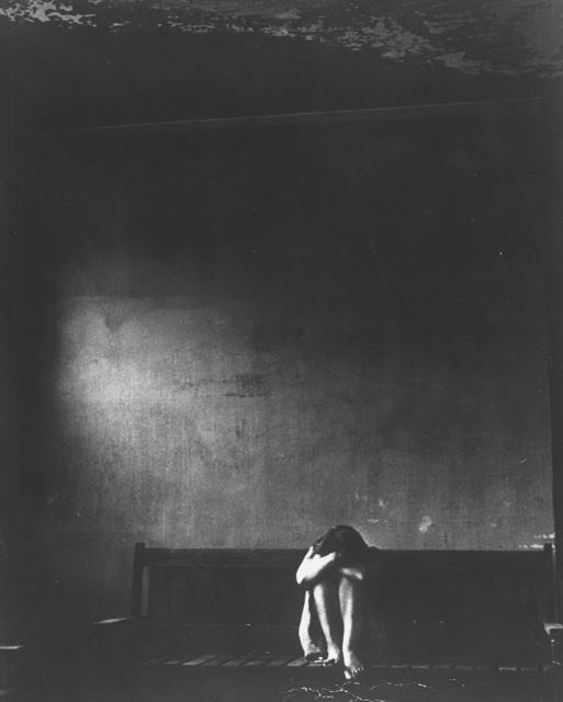
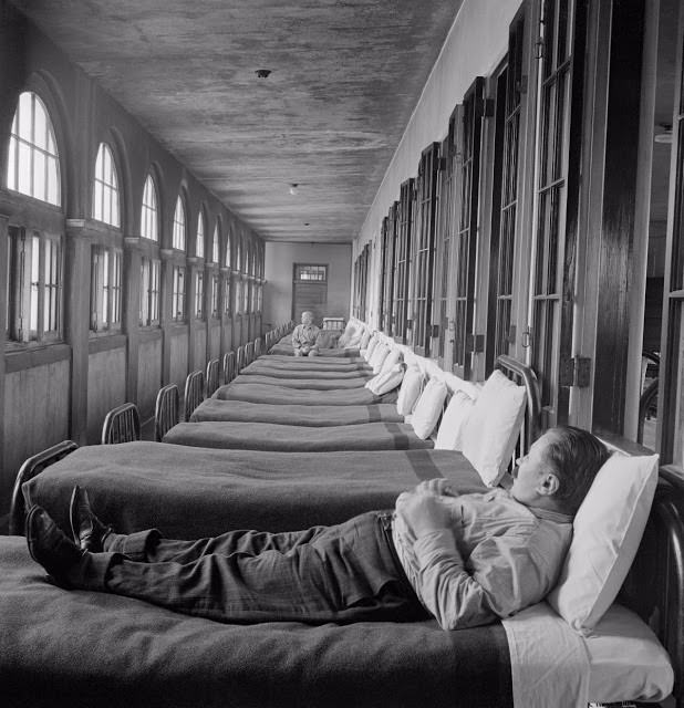

The straightjacket was one of the most widely used restraint tools in psychiatric institutions. Patients were placed in canvas jackets with extra-long sleeves that were tied behind the back, preventing any arm movement. It was used in hospitals across the United States and Europe well into the 20th century, often left on patients for extended periods.
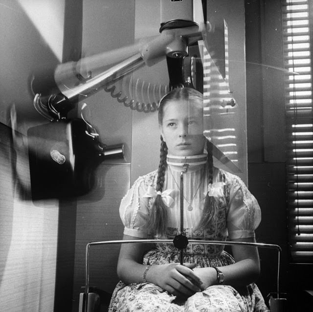
Dental X-rays in the mid-20th century typically required patients to bite down on uncomfortable film plates placed inside the mouth. By 1960, a new machine was developed that circled the entire head, capturing a panoramic image of all the teeth at once without the need for internal film. It was a significant improvement in both patient comfort and diagnostic detail.
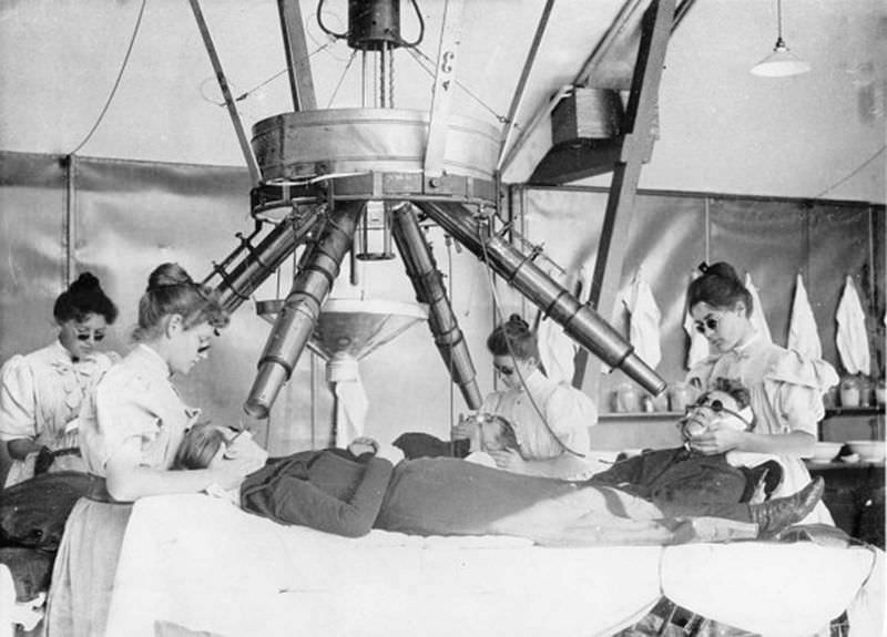
The Institut Finsen in Copenhagen was founded by Niels Finsen, who won the Nobel Prize in Medicine in 1903 for his work using concentrated light radiation to treat lupus vulgaris — a form of skin tuberculosis. Patients at the institute sat in fixed positions while focused beams of light were directed at the affected areas of their skin for long periods.
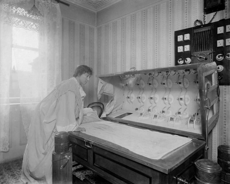
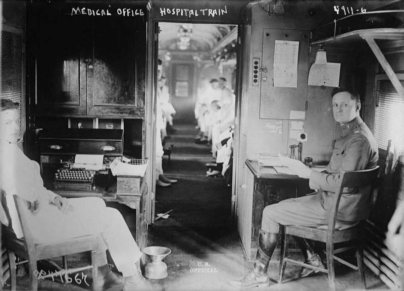
Hospital trains were used to transport wounded soldiers and civilians during conflicts and disasters in the late 19th and early 20th centuries. Medical staff worked in repurposed train cars, performing examinations and basic procedures while in transit. The conditions were cramped and the equipment was limited, but the trains allowed patients to reach larger facilities faster.
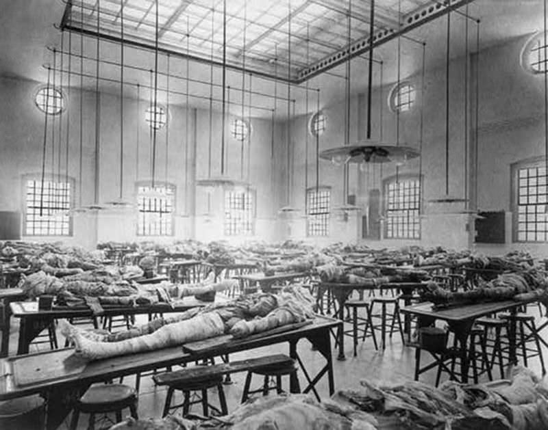
Medical students at the Jefferson Medical College in Philadelphia trained on real human cadavers in a dedicated dissecting room. The practice of cadaver dissection was the primary way medical students learned anatomy. Access to cadavers had historically been so limited — and so controversial — that grave robbing to supply medical schools was common in the 18th and early 19th centuries.
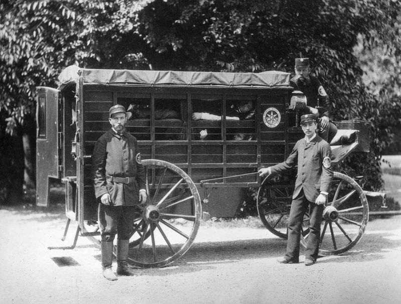
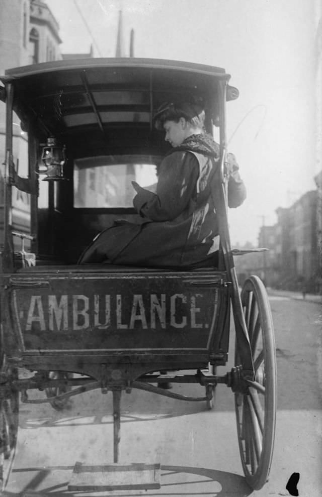
Horse-drawn ambulances were the standard emergency transport in cities across the world until motor vehicles became widely available. Dr. Elizabeth Bruyn operated one in the United States around 1911. Female physicians were rare at that time, and running an ambulance service independently was a particularly unusual role for a woman in early 20th century medicine.
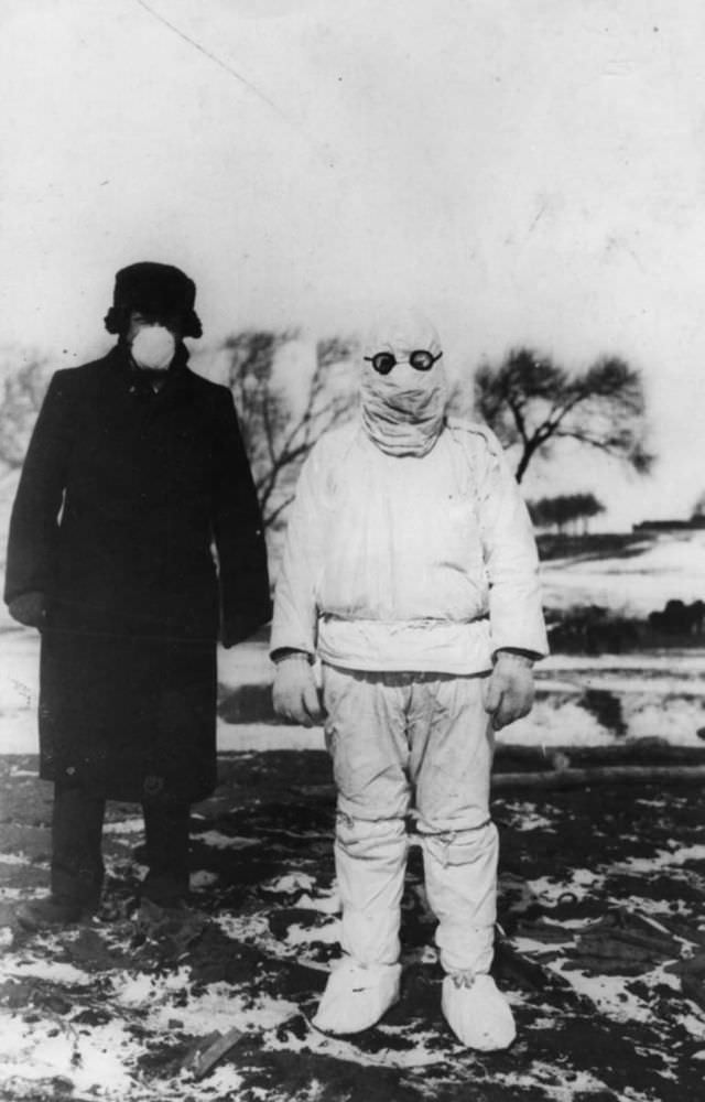
The Manchurian plague epidemic of 1910–1911 killed more than 60,000 people. Doctors treating patients wore early forms of protective suits — heavy robes, gloves, and masks — to reduce the risk of infection. The epidemic was one of the first major outbreaks where germ theory directly shaped how medical workers protected themselves.
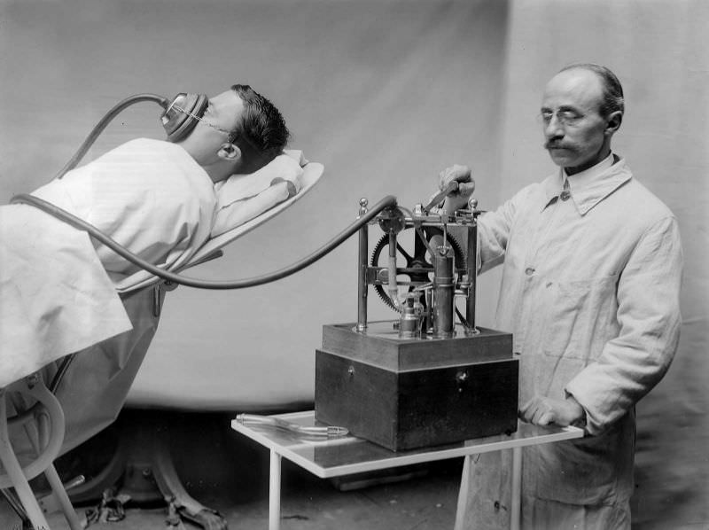
Anesthesia delivery in the early 1910s was still far from standardized. The R. Dubois machine, used in France around 1913, mechanized the process of administering anesthetic gases, allowing for more controlled dosing than older methods where a cloth soaked in ether was simply held over a patient’s face.
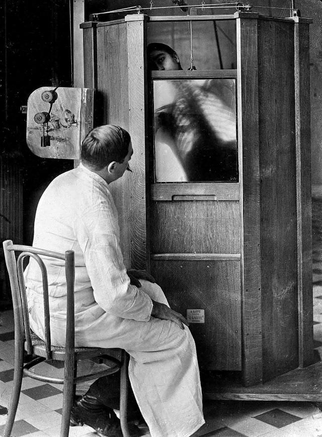
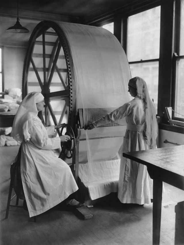
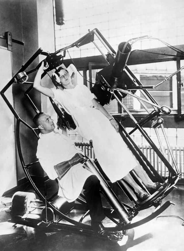
Frankfurt’s 1929 Roentgen machine was specifically designed to protect the operating physician from radiation exposure — a direct response to the growing awareness that earlier X-ray equipment had been injuring and killing the doctors who used it. The machine shielded the radiologist while still allowing full diagnostic imaging of the patient.
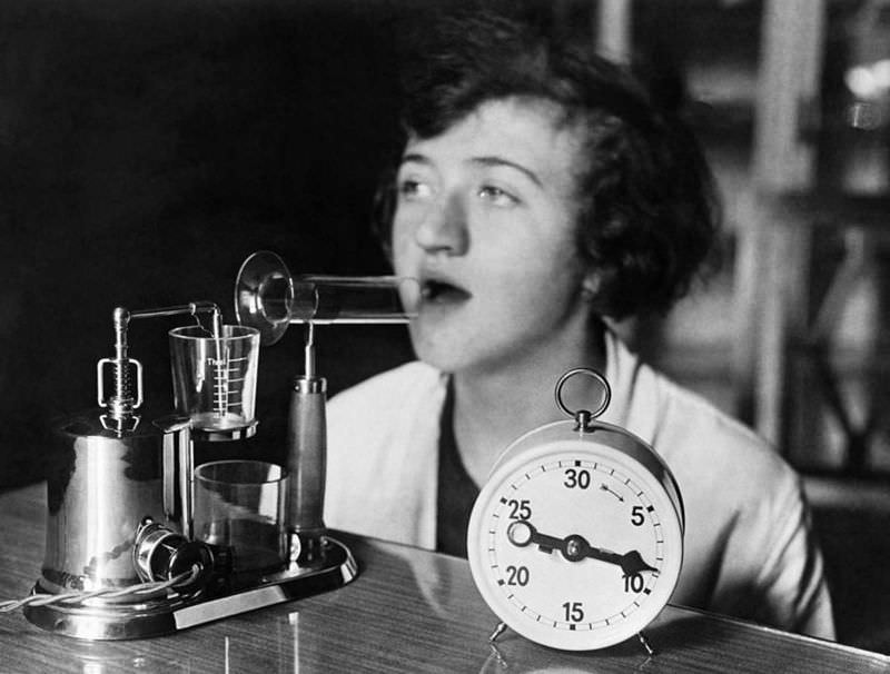
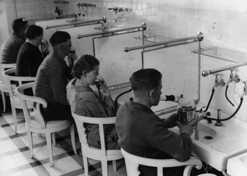
Inhaled medications were used in German hospitals in the 1930s to treat respiratory conditions. Powdered menthol and eucalyptus were delivered directly into the lungs via inhalation devices. The principle was sound — delivering medicine directly to the affected tissue — though the equipment was crude compared to modern nebulizers.
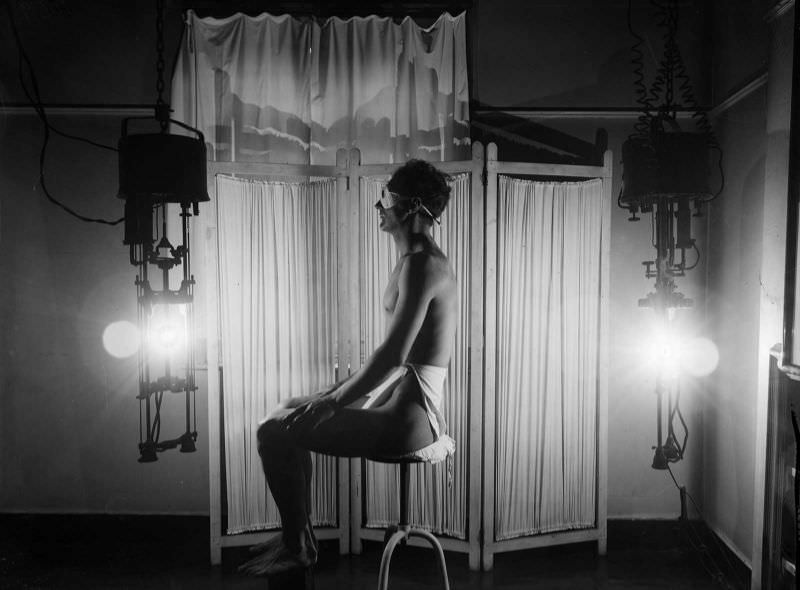
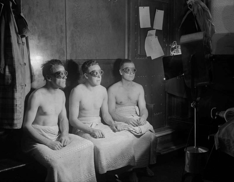
By the early 1930s, UV light therapy had crossed from hospitals into professional sports. Members of the Arsenal Football Club received sunlight treatment in 1931 as part of their physical conditioning. It was thought to boost recovery, improve circulation, and strengthen the immune system.
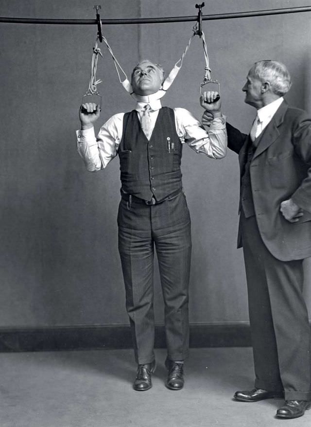
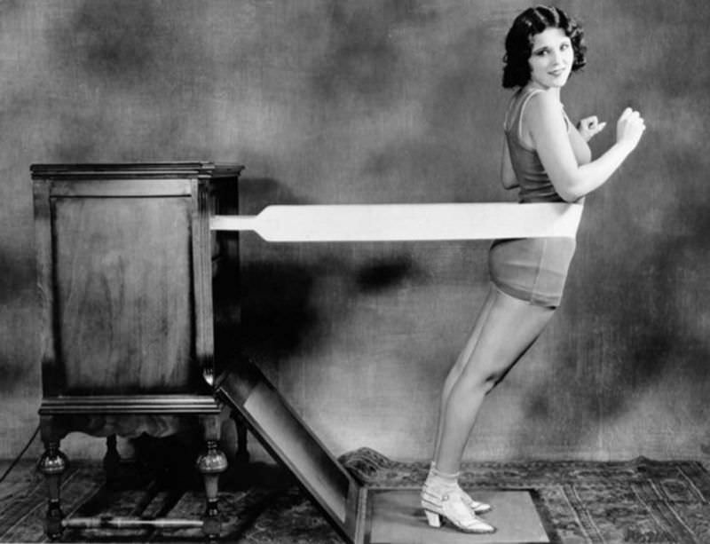
In 1931, a stretching device was marketed with the claim that it could increase a person’s height by two to six inches. A Post Office Department Inspector and a former FDA official tested the device publicly, likely as part of an investigation into fraudulent medical products. Devices making impossible health claims were common during this era, and federal agencies were beginning to push back.
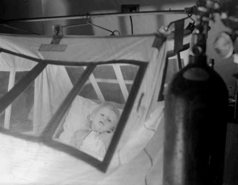
Oxygen tents were used for patients with respiratory conditions who needed supplemental oxygen but were not yet on mechanical ventilation. A clear tent was placed over the patient’s head and upper body and filled with oxygen-enriched air. Young Gerald Blackburn, photographed at Princess Beatrice Hospital around 1937, was one of many children treated this way for conditions like pneumonia or asthma.
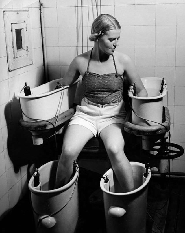
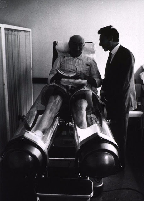
Immersing limbs in water through which an electric current was passed was used in the 1930s to stimulate blood circulation. The patient kept all four limbs submerged simultaneously while current flowed through the water. The treatment was offered in rehabilitation clinics and some hospitals as a response to circulatory disorders.
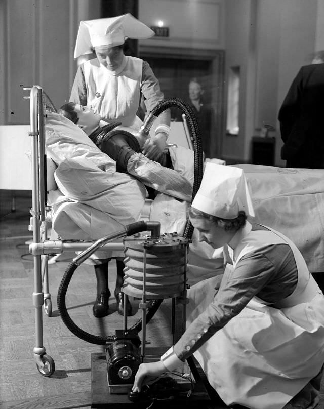
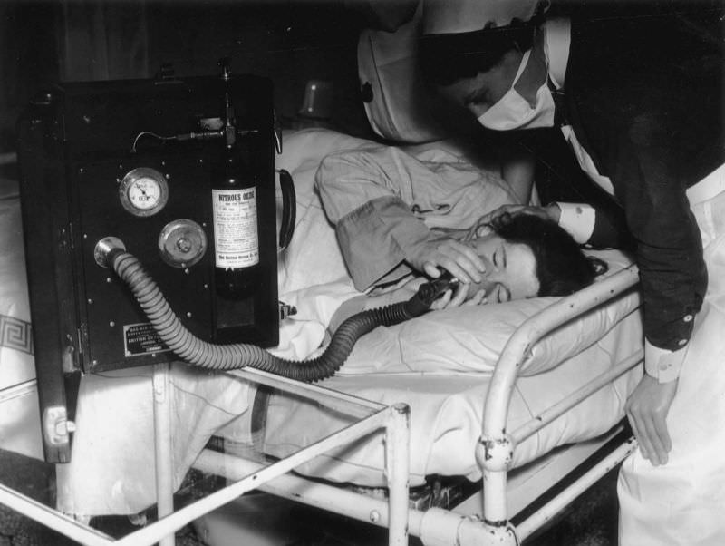
Pain relief during childbirth was limited for most of history. By 1939, inhaled analgesics — typically nitrous oxide or similar gases — were being introduced in some hospitals as a way to reduce pain during labor without fully anesthetizing the mother. A nurse supervised the patient closely to monitor dosage and response.
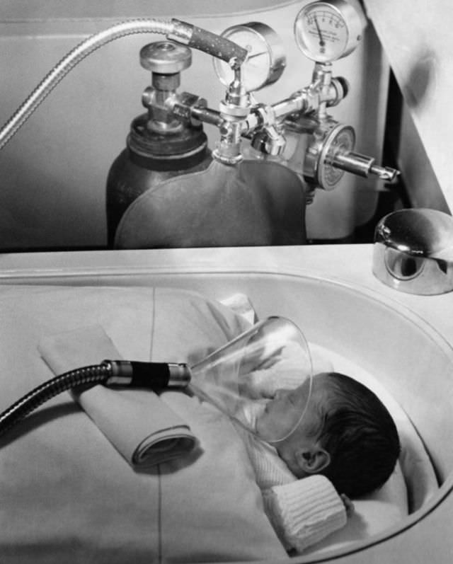
Newborns who struggled to breathe at birth required immediate intervention. In Berlin in July 1939, oxygen was administered to a newborn using equipment that, while basic, represented a meaningful step in neonatal care. Infant mortality from respiratory failure was still extremely high at this time, and the ability to deliver supplemental oxygen to a newborn directly addressed one of its most common causes.

That #8 thing for scoliosis actually worked. They didn’t have the ability to make modern braces so they used those.