The US National Library of Medicine holds millions of books and journals—but tucked inside its vast archive are objects far stranger than any textbook. From gruesome surgical tools to centuries-old illustrations of monsters, the collection reflects how wildly different medicine looked across time. Some items are chilling. Others are just plain bizarre. All of them are real.
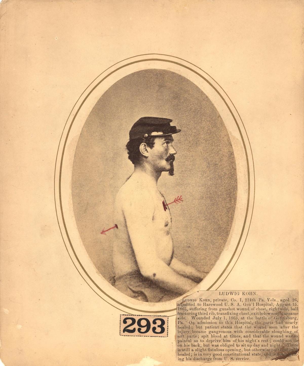
The Civil War-era surgical cards document battlefield medicine in brutal detail. Surgeons at the time worked without modern anesthesia or sterile technique. Amputations were common, and speed was survival. These cards were used by the Army Medical Museum to record and study war wounds—making them some of the earliest examples of medical photography in the United States.
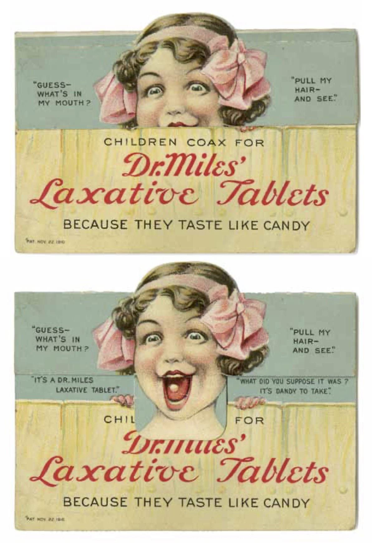
Not everything in the collection is grim. Around 1910, pharmaceutical companies used clever advertising tricks to sell their products. This die-cut novelty card for Dr. Miles’ Laxative Tablets features moving parts—a surprisingly playful approach to marketing medicine. It sits alongside more serious artifacts, showing that even medical history has a quirky side.
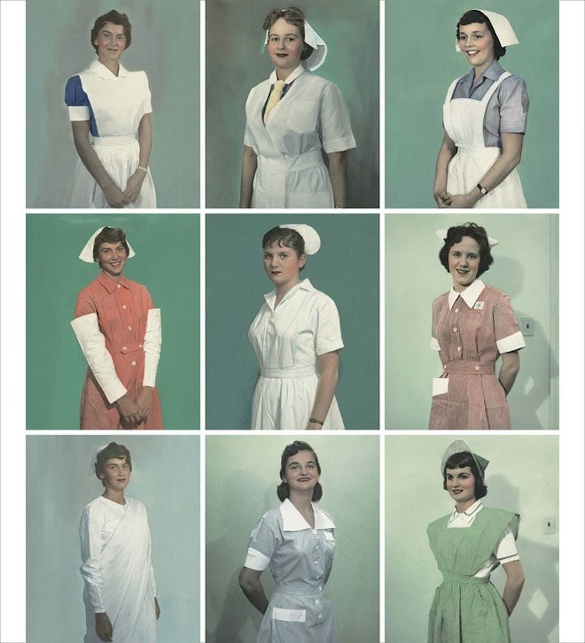
The library also preserves cultural snapshots of healthcare workers around the world. This photograph collection documents nurse uniforms from different countries around 1950, capturing how professional identity in medicine varied by nation and tradition.
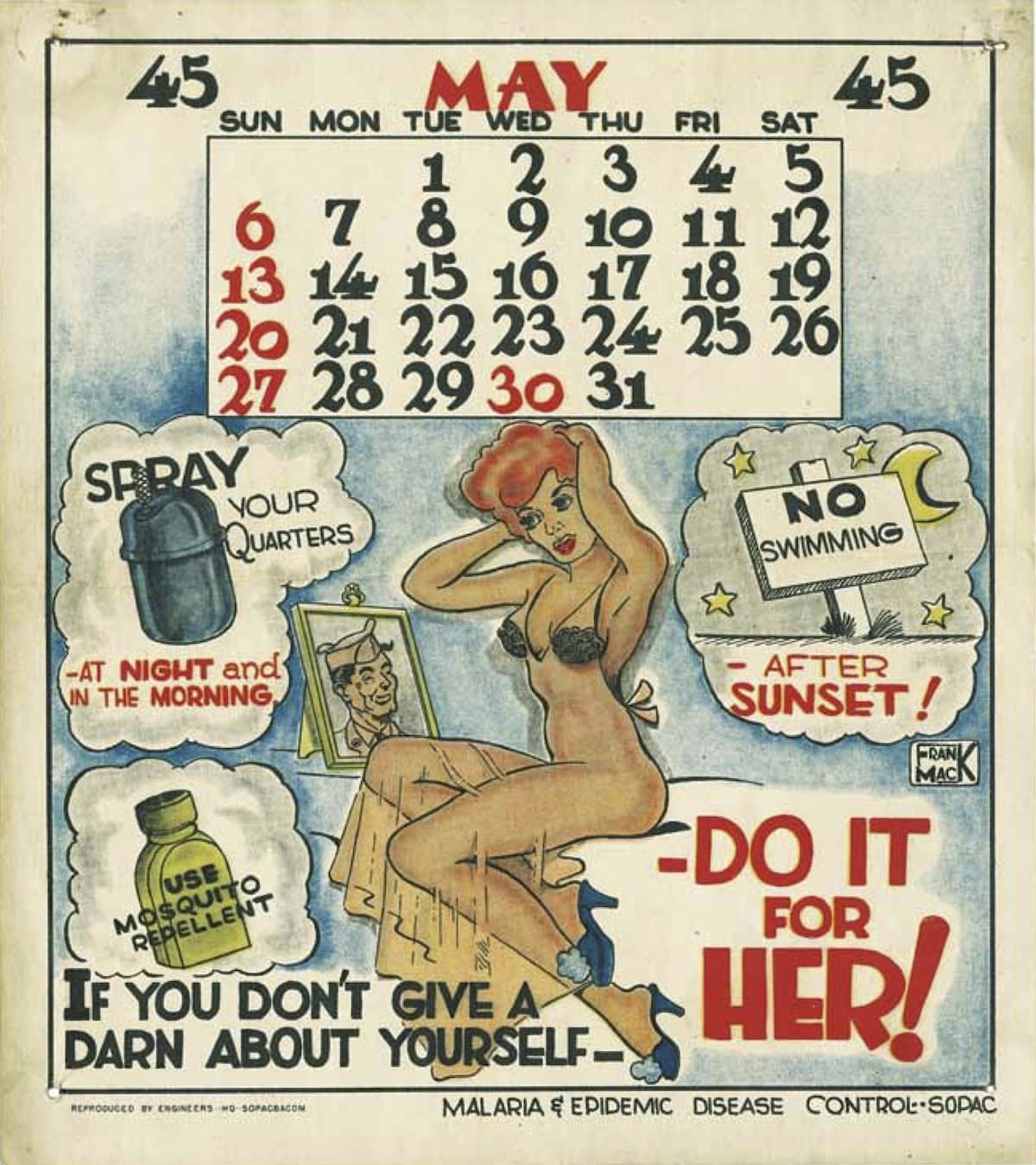
During World War II, the U.S. Army used unconventional methods to get soldiers to take malaria prevention seriously. Frank Mack illustrated pinup-style calendars in 1945 to promote anti-malaria behavior. It was public health messaging designed to grab attention in a military setting—and it worked by meeting soldiers where they were.
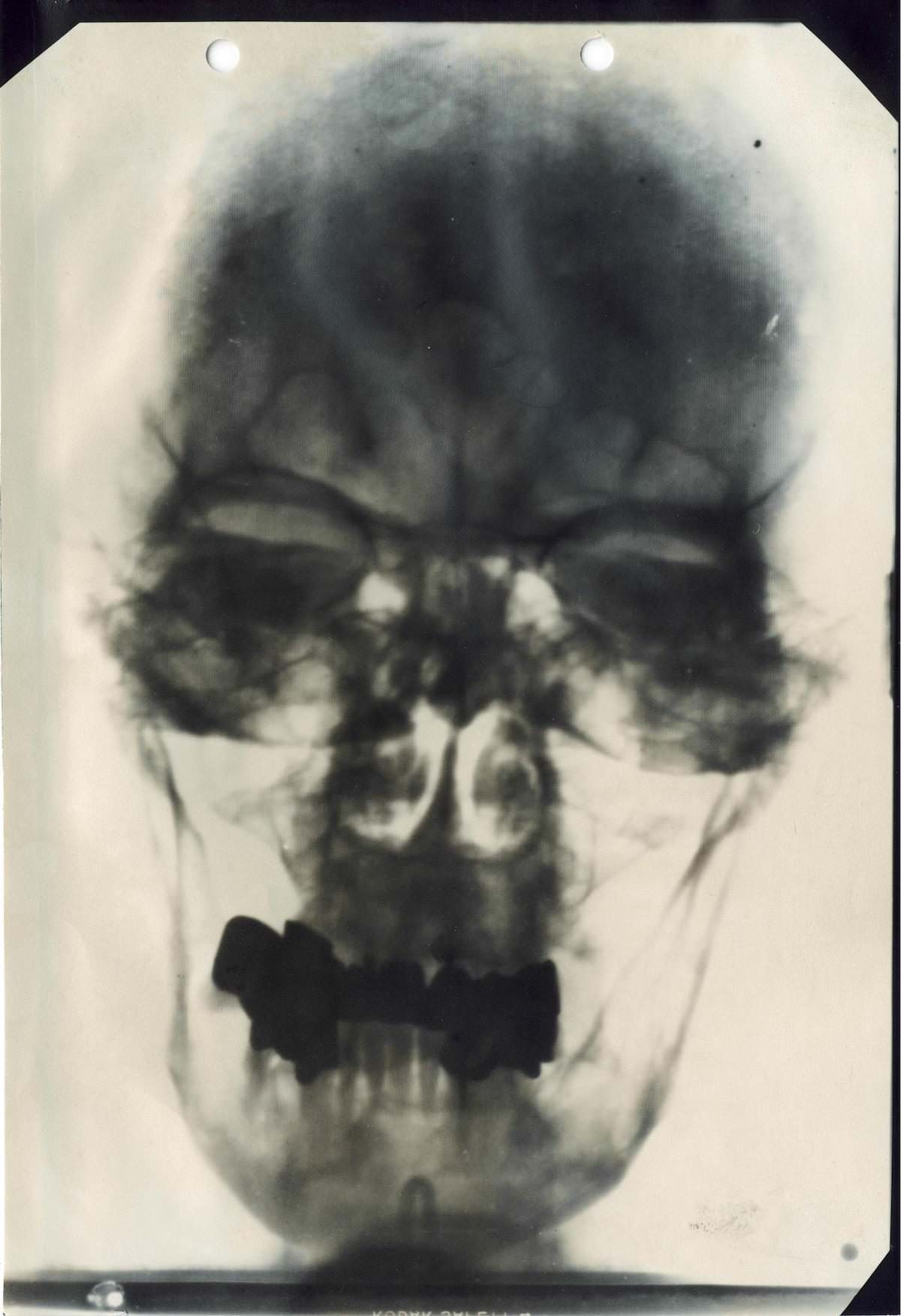
One of the most historically significant items is an X-ray of Adolf Hitler’s nasal passages and teeth, taken by his personal physicians. The document comes from a file titled ‘Hitler as Seen by His Doctors,’ compiled after World War II. These medical records were used by Allied investigators to confirm Hitler’s identity and study his health history.
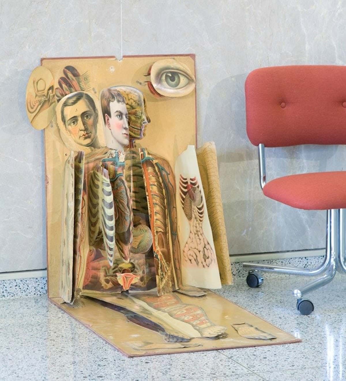
Before digital anatomy tools, medical students relied on physical models like this physiological manikin from 1886. Published by James T. White & Co., it used layered paper illustrations to show internal organs and body systems. It was an affordable, portable teaching tool at a time when cadavers were scarce and anatomy books were expensive.
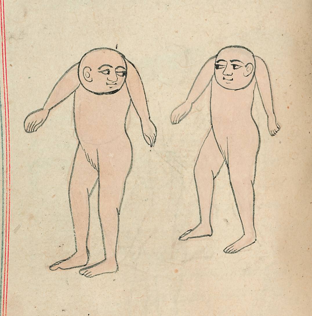
Dating back to the mid-1200s, this cosmographical work by al-Qazwini blends natural history, medicine, and myth. It describes creatures, plants, and phenomena from across the known world—some real, some fantastical. The library holds it as a record of how medieval Islamic scholars understood the natural world and the human body within it.
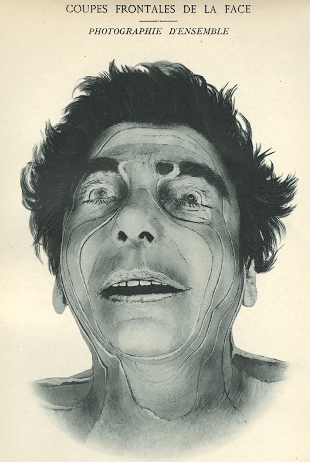
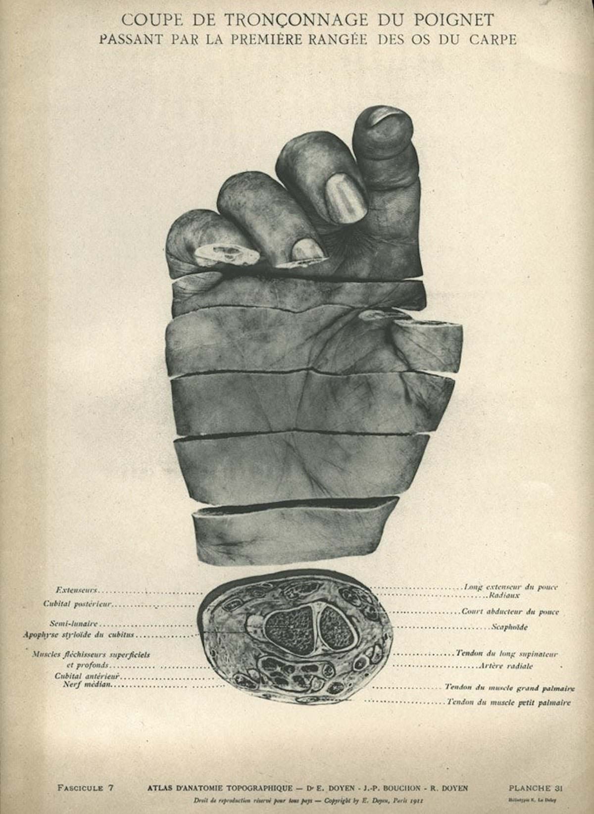
Surgeon Eugène-Louis Doyen took anatomical illustration to a new level in 1911 with this atlas of topographic anatomy. Using photography rather than hand-drawn plates, the work documented the precise locations of muscles, vessels, and organs. It was a landmark shift in how surgical education used visual media.
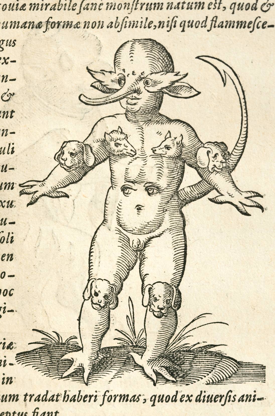
In the 16th century, unusual births were often interpreted as divine signs or warnings. The ‘Monster of Crackow,’ reported in 1547, was described as a child born with claws, a tail, animal heads at the joints, and eyes on its stomach. This illustration from ‘De conceptu’ shows how early modern Europeans mixed medicine, religion, and fear when confronting birth anomalies they could not explain.
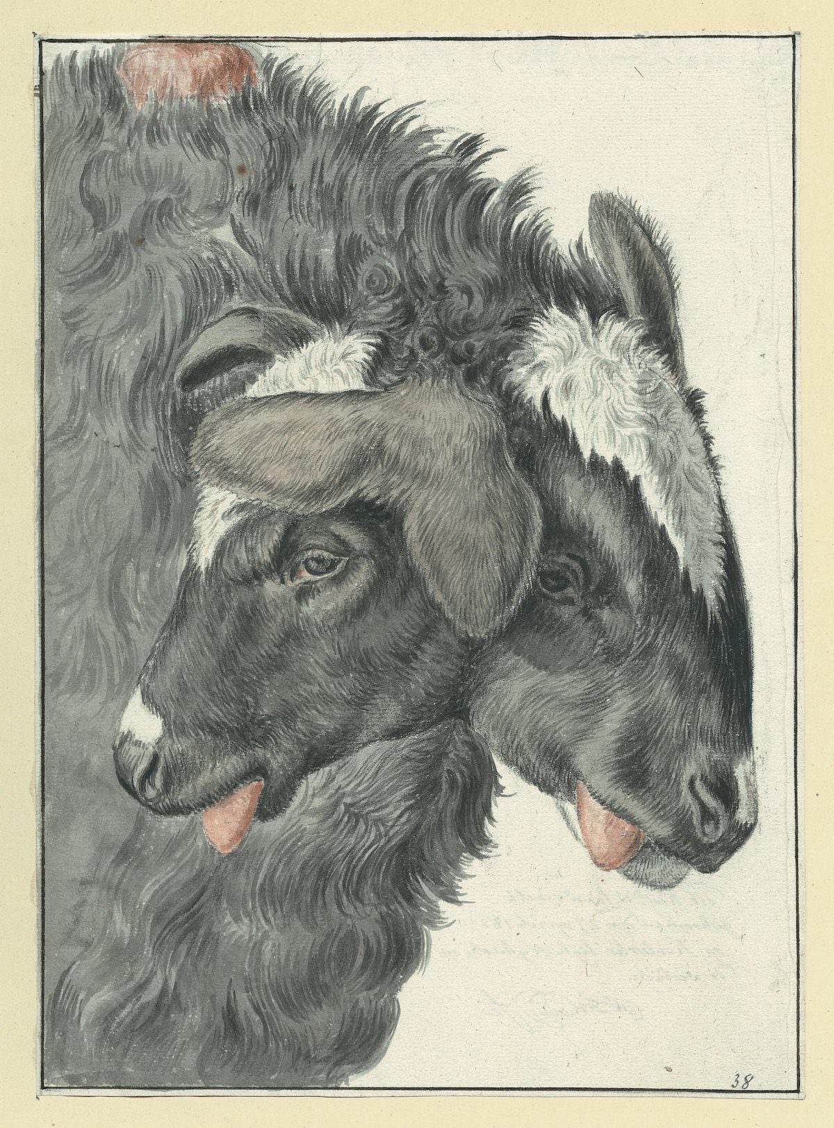
Published in 1587, Jakob Rueff’s work on human conception and generation was one of the first books to systematically address reproductive medicine. Translated by Wolfgang Haller, it combined practical obstetrical knowledge with illustrations that reflected the anatomical understanding of the era—some accurate, others shaped by superstition.
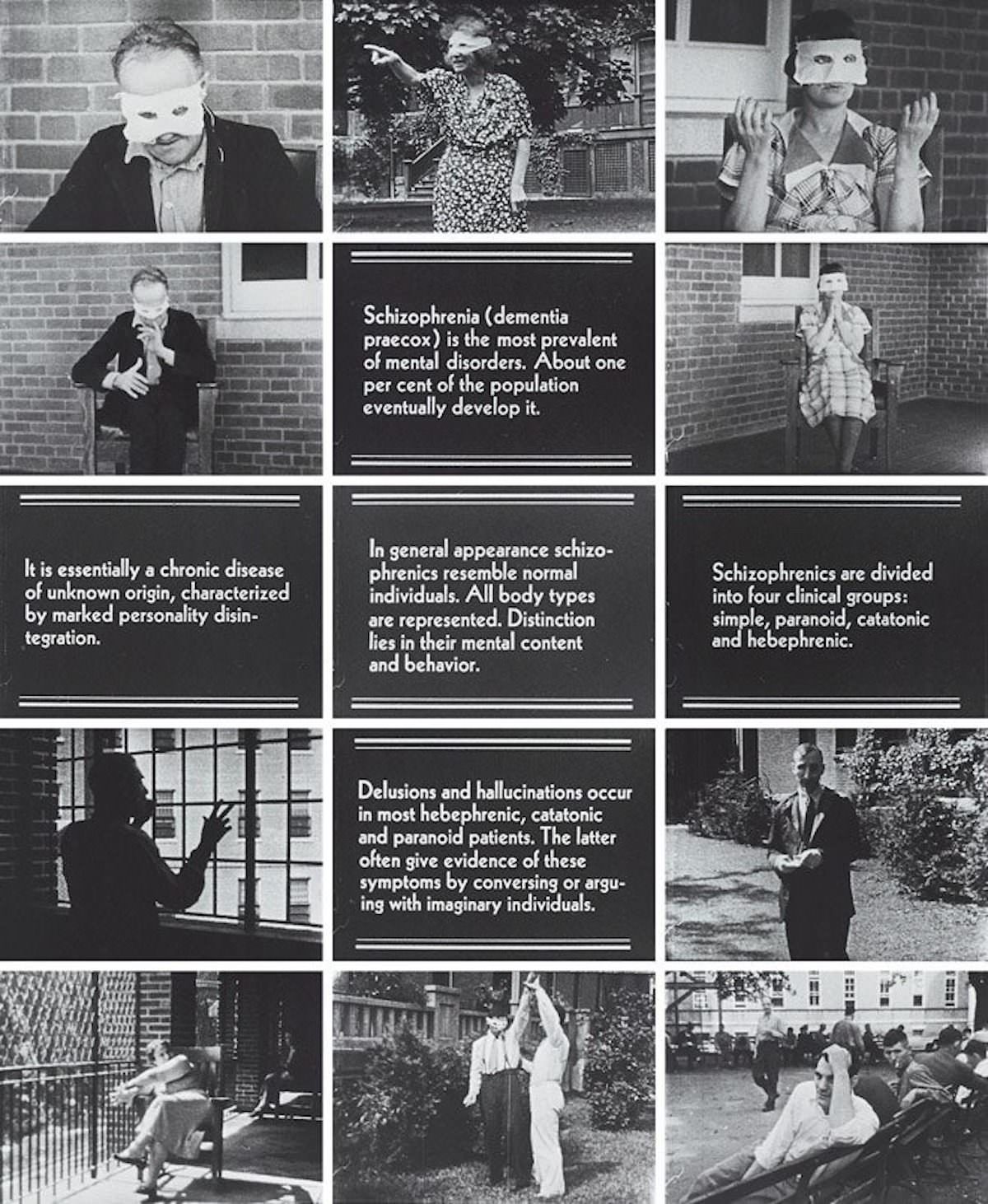
Psychiatric medicine in the early 20th century was still finding its footing. James D. Page’s work on schizophrenia symptoms from around 1930 used illustrated and photographic documentation to categorize patient behaviors. The library holds it as a record of how psychiatry developed its diagnostic frameworks during a critical period.
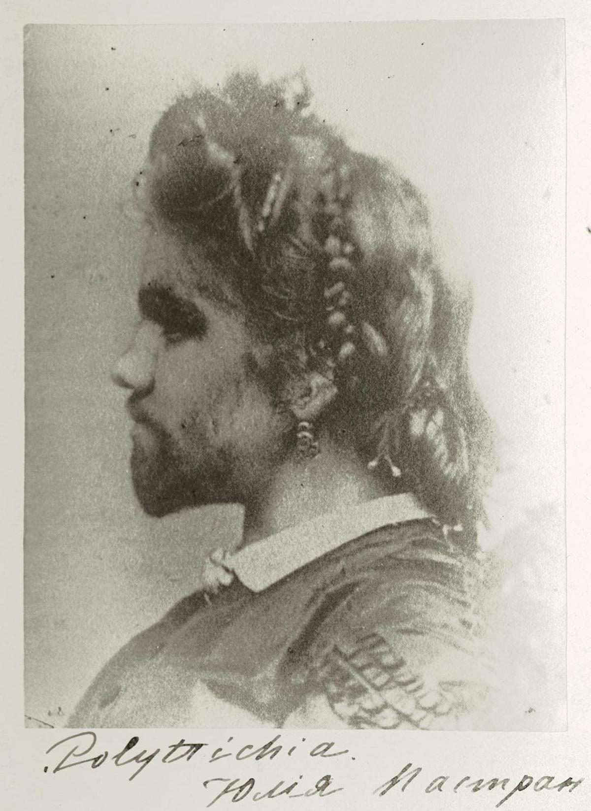
Julia Pastrana was a Mexican performer known as ‘The Bearded Lady’ who toured Europe and North America in the 1850s. She had hypertrichosis, a condition causing hair growth across the face and body. Pastrana died in Moscow in 1860 during childbirth. Her story is preserved in medical literature as both a clinical case and a record of how 19th-century medicine and entertainment intersected—often exploitatively.
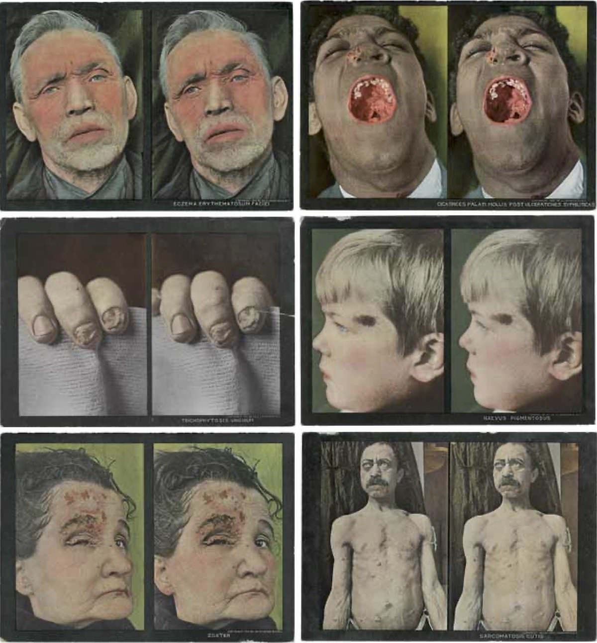
In 1910, Selden Irwin Rainforth used stereoscopic photography to document skin diseases in three dimensions. The format allowed physicians to study lesions and conditions with more depth than flat photographs could provide. It was an early example of technology being adapted specifically for medical education.
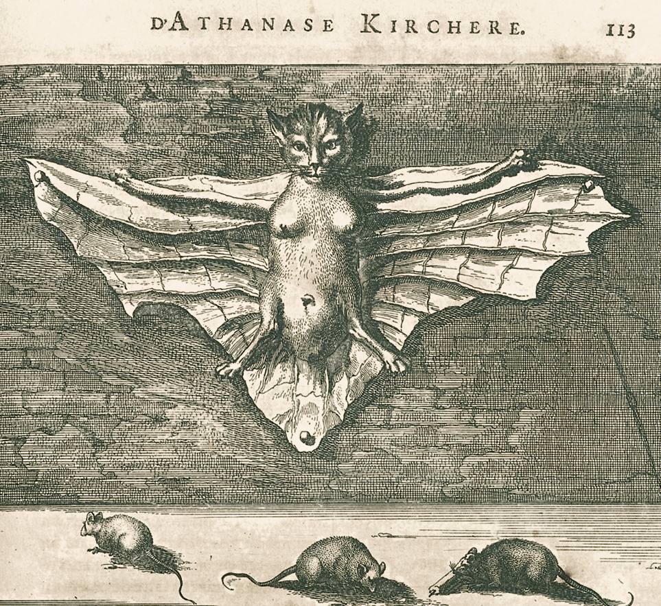
Athanasius Kircher was a 17th-century Jesuit scholar who wrote extensively on natural history, China, and the occult. This illustration from ‘La Chine’ depicts a cat with a female torso and bat wings nailed to a wall above feeding mice—an image that reflects the era’s blend of zoological curiosity and supernatural thinking. The library holds Kircher’s works as examples of pre-scientific natural philosophy.
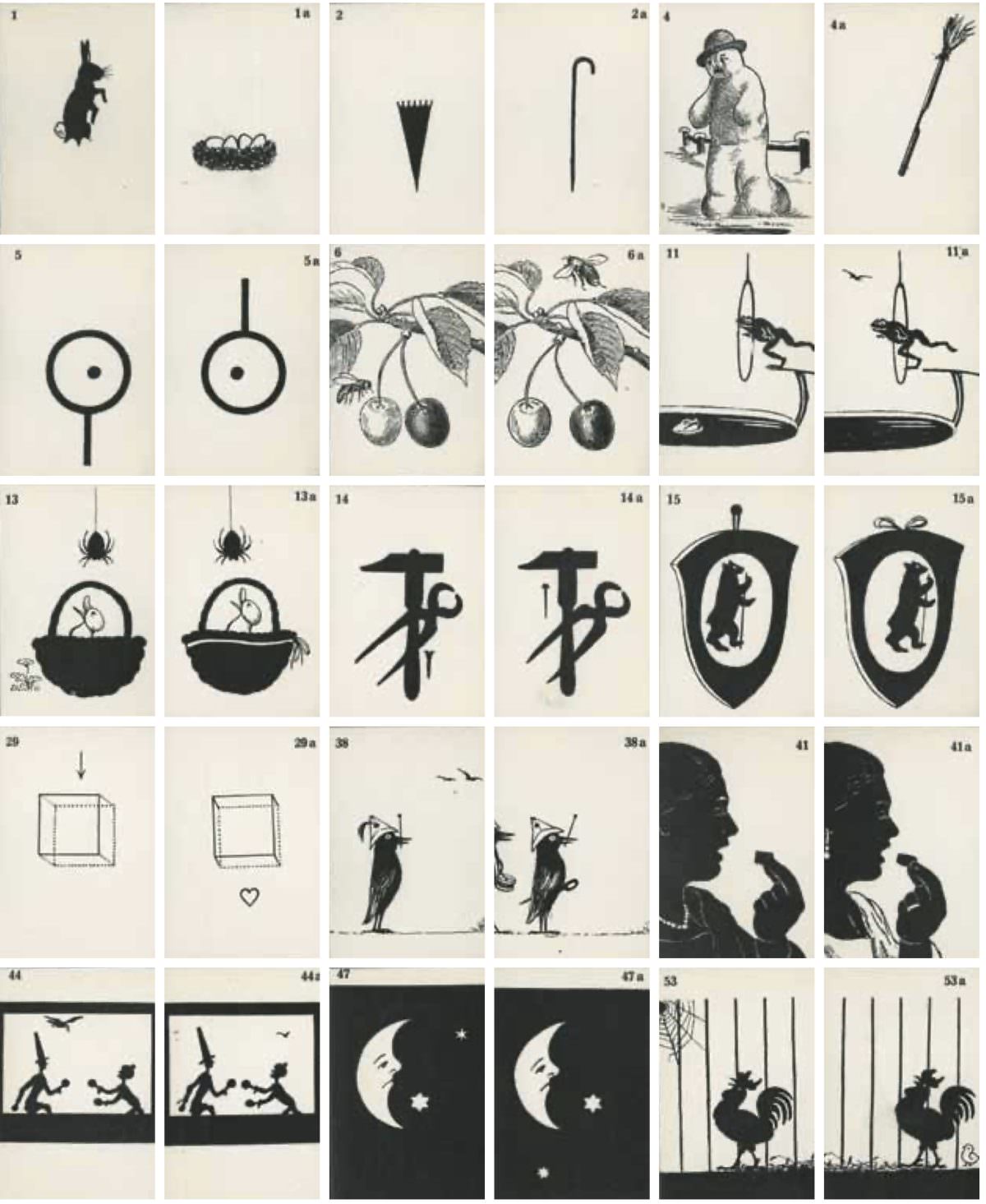
Carl Hubert Sattler developed stereoscopic images in 1942 as a treatment tool for children with strabismus, commonly called crossed eyes. The visual exercises were designed to train the eyes to work together. It’s a small but specific example of how vision medicine used emerging optical technology for therapeutic purposes.
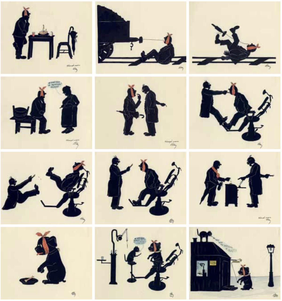
Otto Elkan produced a series of dental cartoons around 1945, using humor to communicate oral health messages. Like the Army’s malaria pinups, these cartoons show that medical communicators understood the power of accessible, entertaining formats to reach the public—especially during and after wartime.
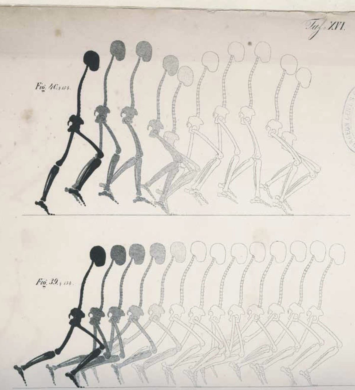
Brothers Wilhelm and Eduard Weber published a detailed mechanical analysis of human walking in 1836. Using physics and measurement, they broke down gait into precise movements—one of the first scientific studies of human locomotion. The library holds it as a foundational text in biomechanics.
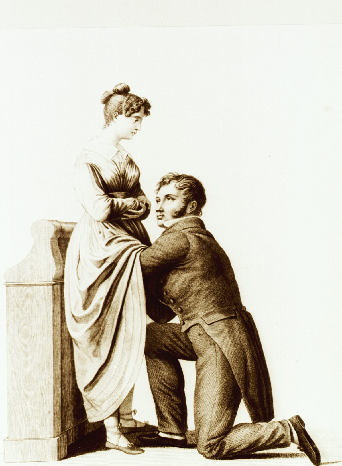
Jacques Pierre Maygrier’s 1822 obstetrical atlas from Paris contains detailed illustrations of childbirth procedures and examinations. At the time, obstetrics was becoming a formal medical specialty, and visual guides like this one trained physicians in techniques that had previously been passed down informally or left entirely to midwives.
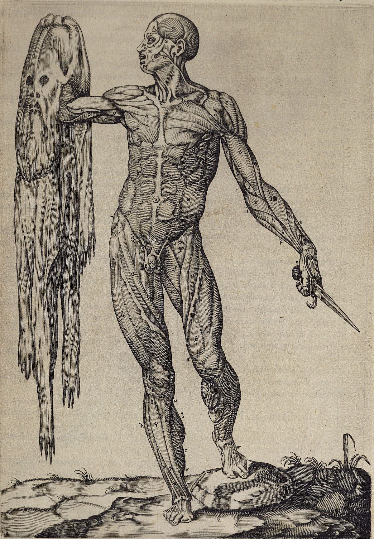
Juan Valverde de Amusco’s 1550 anatomy text includes one of the most striking images in medical history: a flayed cadaver holding its own skin in one hand and a dissecting knife in the other. Published in Rome, the illustration was both educational and theatrical—designed to impress as much as inform. It remains one of the most reproduced images from Renaissance anatomy.
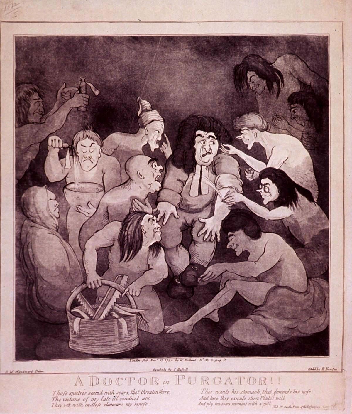
Satirical cartoons about doctors have a long history. George Moutard Woodward, working around 1760–1809, produced caricatures that mocked medical practitioners and their treatments. His work reflects widespread public skepticism of physicians during a period when medical practice was often more harmful than helpful.
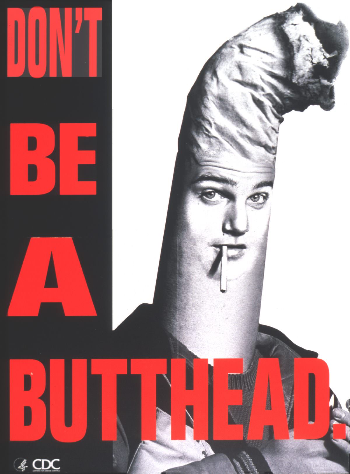
From the CDC in 1994, this anti-smoking poster takes a blunt, irreverent approach to public health messaging. ‘Don’t be a butthead’ was aimed at younger audiences at a time when tobacco companies were aggressively targeting teens. The library preserves it as part of its collection of public health communication materials.
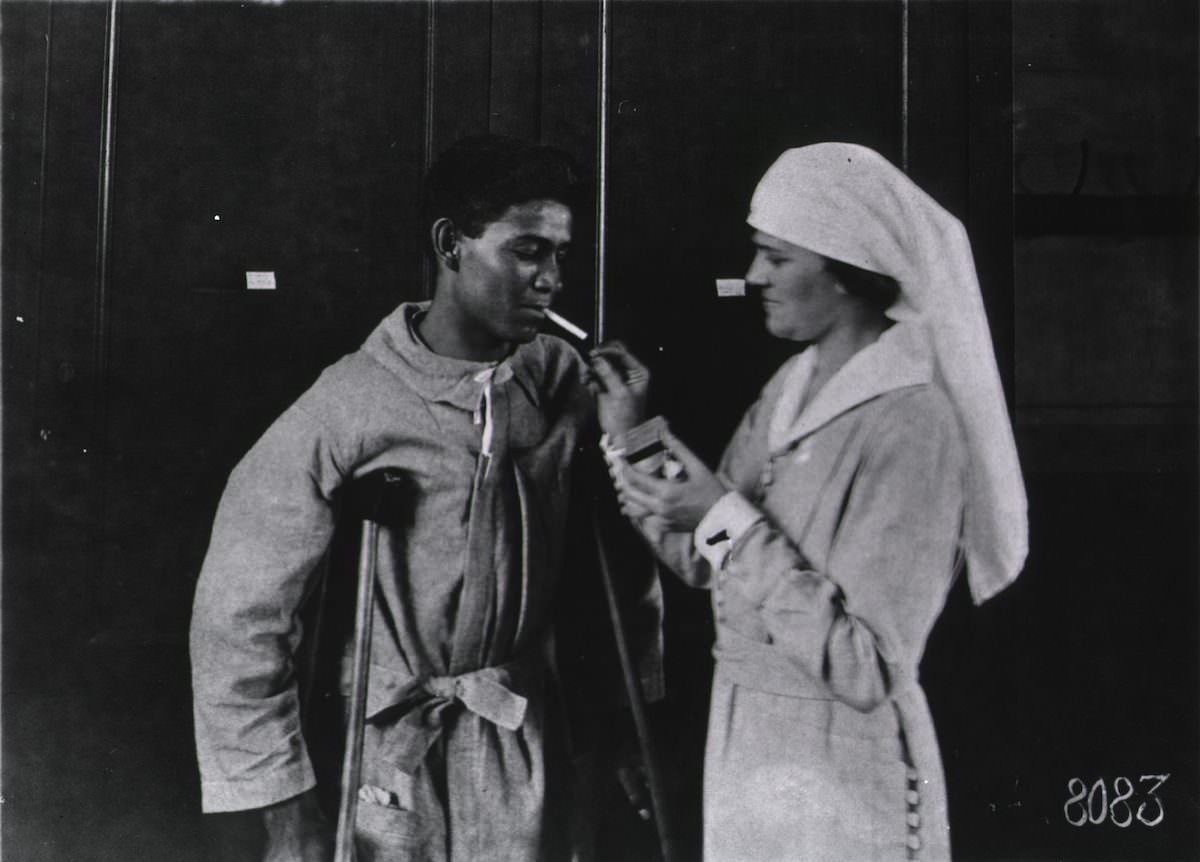
This photograph from U.S. Army Base Hospital Number 41 in Paris documents a Mexican soldier being treated while fighting for America. It’s a rare visual record of the diverse men who served in and were cared for by American military medicine—a part of the historical record that has often been overlooked.