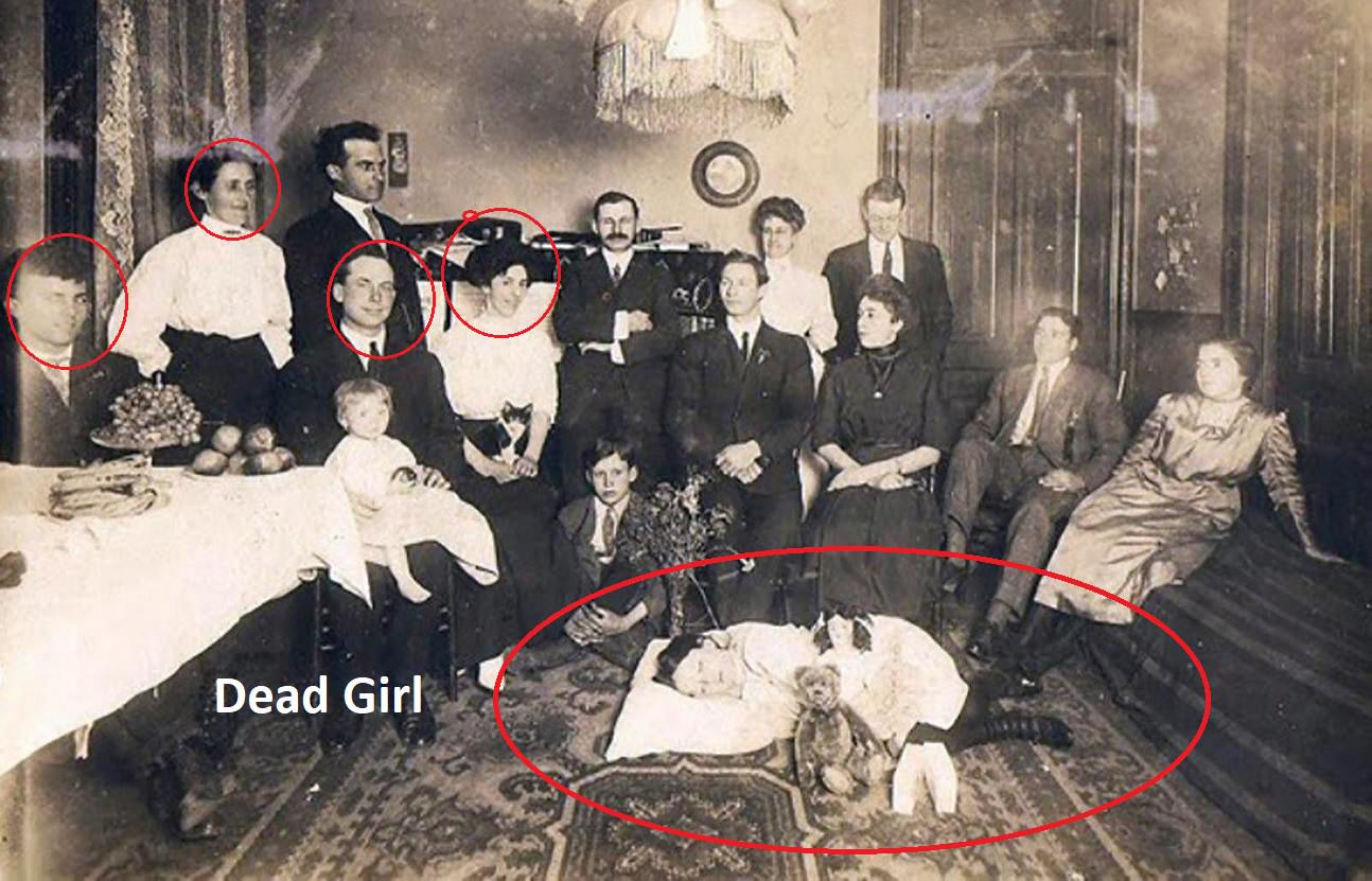
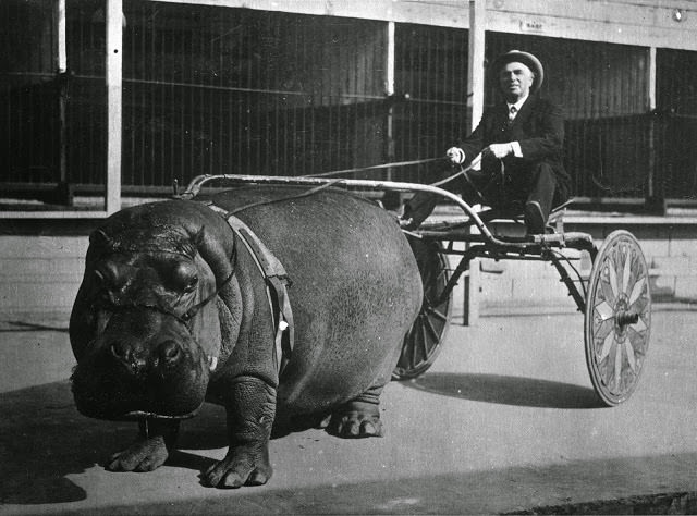
In the 19th century, death was not hidden away. Photographing dead family members was a common practice called post-mortem photography. Cameras were expensive and rare, so a post-mortem portrait was often the only photograph a family ever had of a person. The deceased were carefully posed to look as though they were simply sleeping.
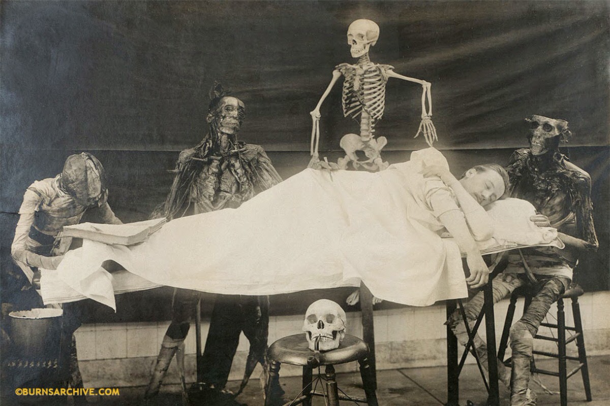
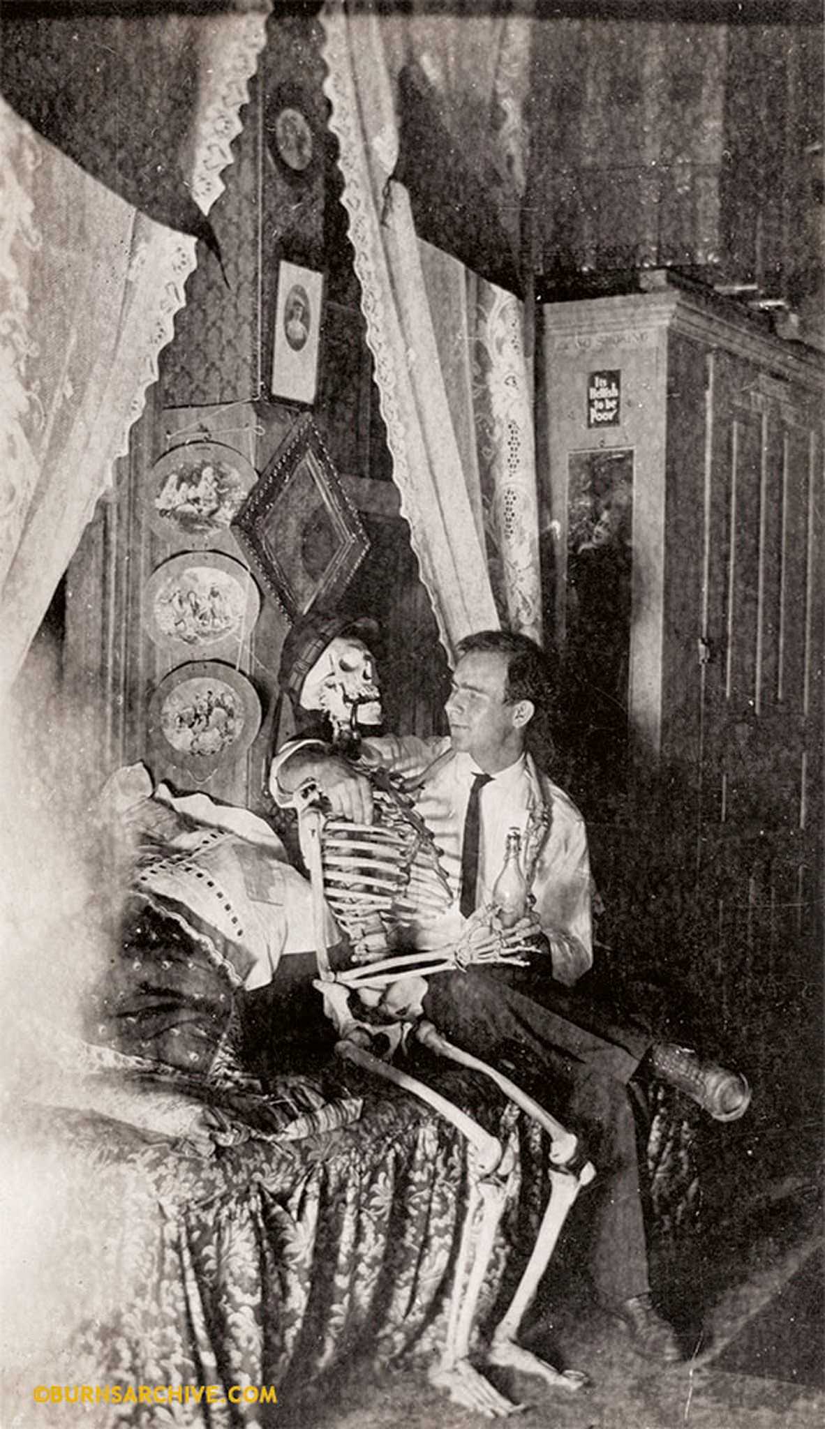
At the same time, medical photography was growing fast. Doctors and scientists used cameras to document the human body in precise detail. Medical schools became a common setting for these images, where students posed alongside skeletons as part of their anatomy lessons.
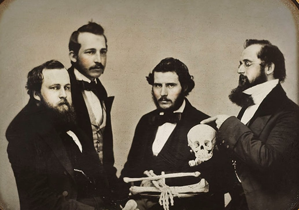
The Burns Archive holds hundreds of these early photographs. They show a deep scientific curiosity about bones and body parts that was entirely normal for the era. Skeletons were sometimes dressed in clothing or arranged in lifelike poses, reflecting a comfort with human remains that modern culture no longer shares.
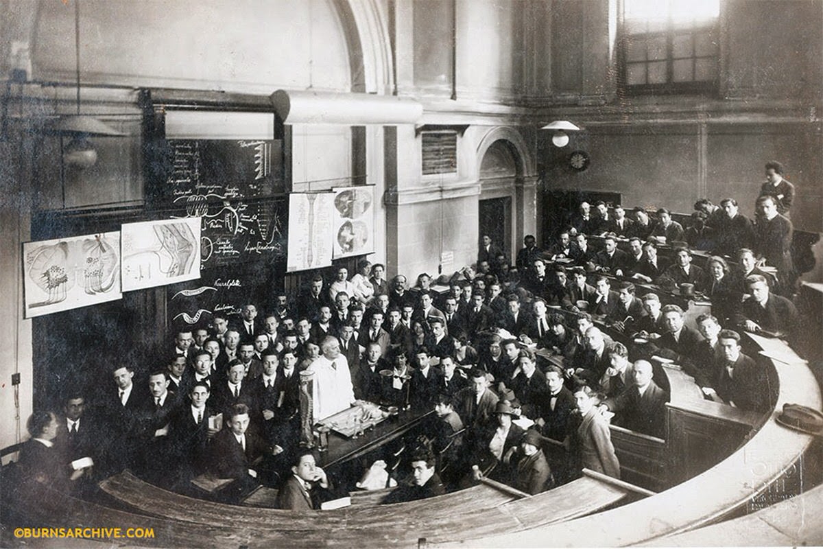
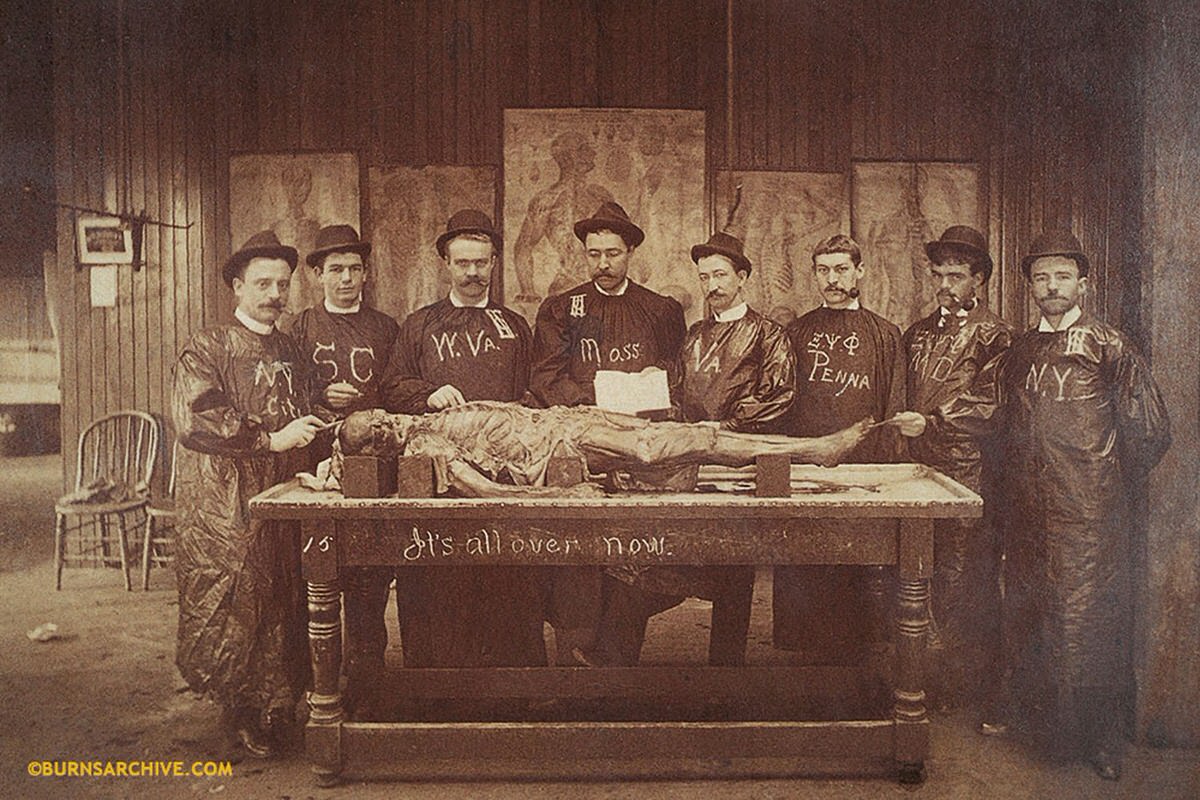
By the early 20th century, as Dr. Stanley B. Burns explains, death moved out of the home and into hospitals and funeral parlors. Dead bodies disappeared from personal photographs along with it. Death became a private event rather than a family or public one.
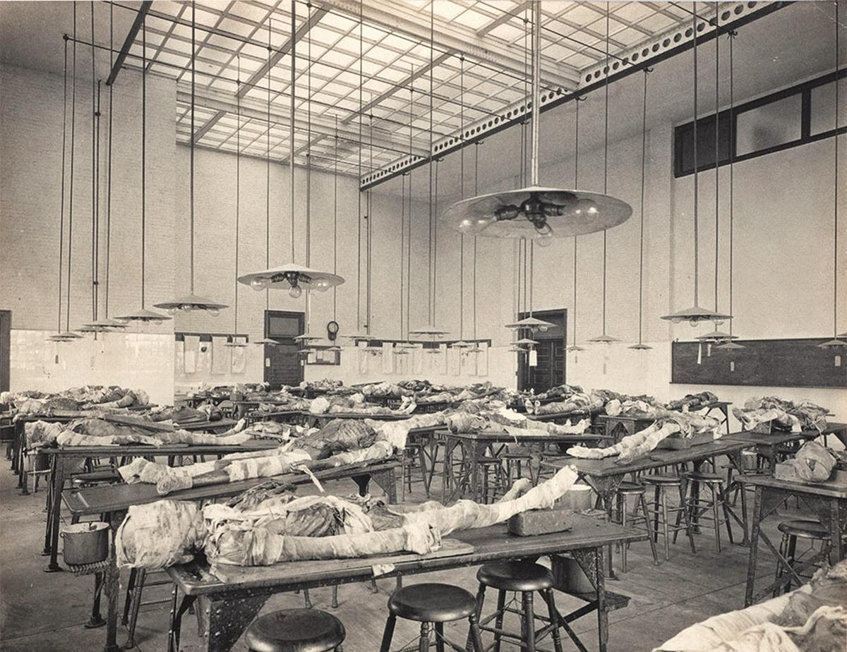
The book Stiffs, Skulls & Skeletons collects 450 photographs from this period, covering everything from medical oddities to formal skeletal portraits. The early black-and-white prints capture bone and skin in striking detail.
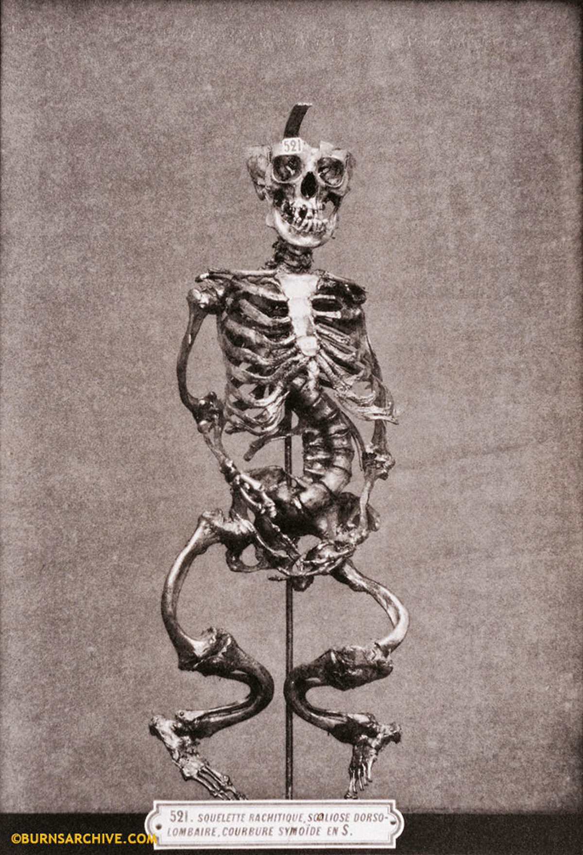
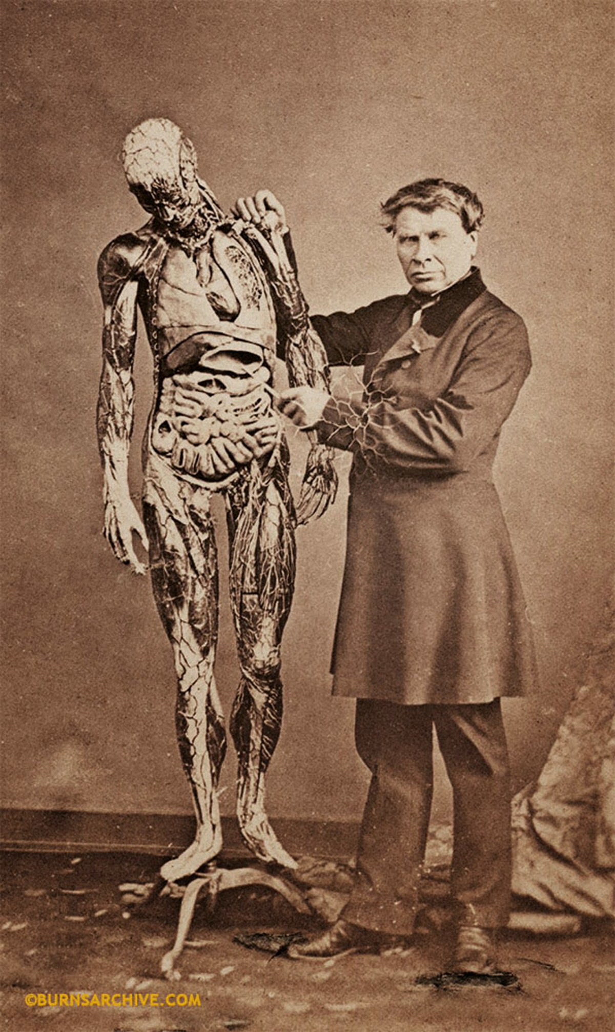
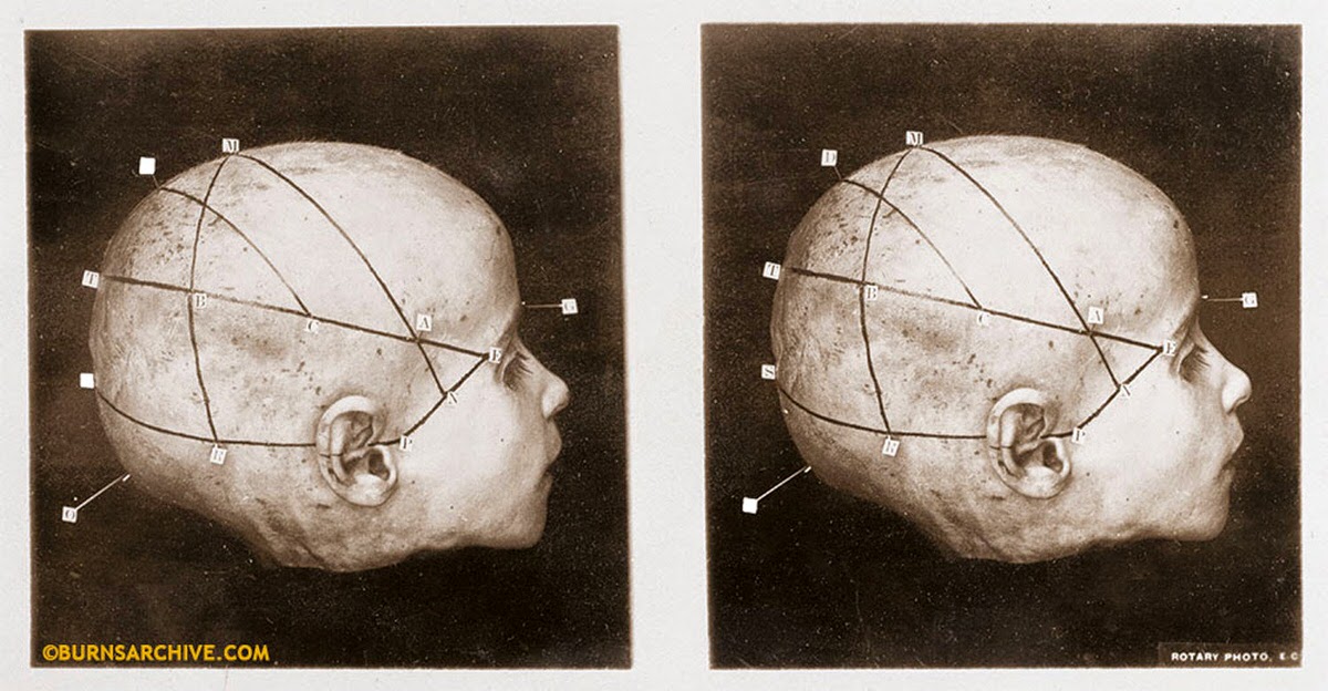
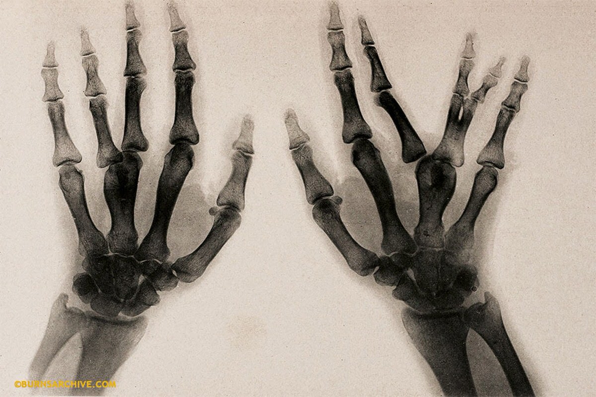
These photographs also served hard scientific purposes. Doctors recorded skeletal deformities, mapped brain anatomy onto the skull, and used X-rays to study abnormal bone structures — all documented with a camera.
Not every skeleton photo was purely academic. Some were sent as novelty postcards between friends and neighbors, treating human remains as a lighthearted curiosity rather than a source of dread.