
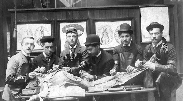
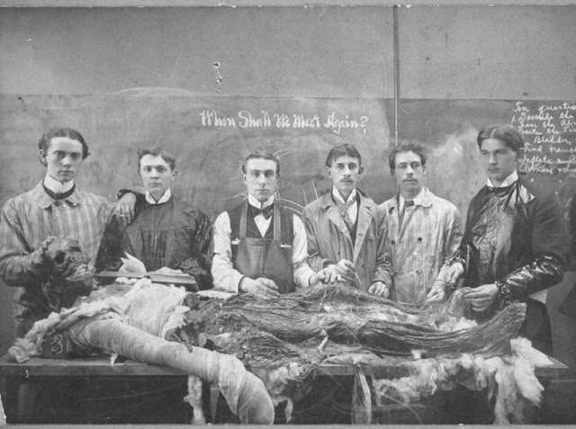
Long before sterile gloves, digital scans, and plastic models, medical students learned anatomy the hard way — with bare hands and a human body on the table. The Victorian era, roughly spanning the early 1800s through the end of the 19th century, produced some of the most intense and revealing moments in the history of medical education. Students crowded around cadavers in cold, poorly lit dissection rooms, cutting through flesh and bone to understand what made the human body work. These photographs capture that world.
The roots of anatomy go back to around 300 BCE in Alexandria, where physicians dissected human corpses to study disease. Ancient Greek and Roman scholars built on that work, creating the first written records of how the body was structured. But after the Roman Empire collapsed and the Christian Church gained power across Europe, human dissection was widely condemned. That prohibition lasted for centuries and pushed anatomical study into the shadows.
The Renaissance cracked the door back open. Scholars driven by intellectual ambition began challenging religious restrictions, carefully and quietly at first. Leonardo da Vinci dissected dozens of human bodies and filled notebooks with precise anatomical drawings. Andreas Vesalius, a Flemish physician working in the 1500s, went further — he performed public dissections and published detailed illustrated texts that corrected centuries of errors. Their work was groundbreaking, but it still existed mostly within narrow academic circles. The general public remained deeply uncomfortable with the practice.
By the late 18th century, medicine was transforming into a respected profession, and dissection was increasingly seen as essential training. New medical schools opened across England, and enrollment surged. Anatomy became a core part of the curriculum. The problem was simple and grim: there were not enough bodies.
English law at the time permitted medical schools to use the bodies of executed criminals. The Murder Act of 1752 had formally established this practice, framing dissection as an additional punishment on top of execution — a deterrent meant to horrify the public. But executions could not supply nearly enough cadavers to meet the demand from a growing number of medical schools and their students.
The shortage created a black market. Grave robbers known as “Resurrectionists” began digging up freshly buried bodies at night and selling them to anatomy professors and medical schools. It was illegal, deeply offensive to grieving families, and widely despised — but it was also lucrative. Schools paid well, and demand never dried up. The trade was open enough that many people in the medical community knew exactly where their cadavers came from.
Families of the recently buried fought back. Wealthy households purchased iron coffins that were nearly impossible to pry open. Some cemeteries installed stone vaults over fresh graves. Others hired armed watchmen to guard burial grounds through the night. Certain graveyards even rigged gunpowder traps to deter nighttime intruders. Despite all of this, the Resurrectionists kept working. The money was too good and the schools too desperate.
The worst known case tied to this era involved not grave robbing but murder. In Edinburgh, William Burke and William Hare killed at least 16 people between 1827 and 1828, selling the bodies directly to the anatomist Robert Knox. When they were caught and Burke was tried and executed in 1829, public outrage reached a breaking point. The case made it impossible for Parliament to ignore the connection between anatomy’s demand for bodies and the crimes being committed to supply them.
Parliament responded with the Anatomy Act of 1832. The law overhauled how cadavers were obtained for medical education. It allowed the bodies of people who died in workhouses or hospitals and went unclaimed to be used for dissection. It also opened the door for individuals and families to donate bodies voluntarily. The act effectively ended the legal need for body snatching and removed the criminal market that had sustained it.
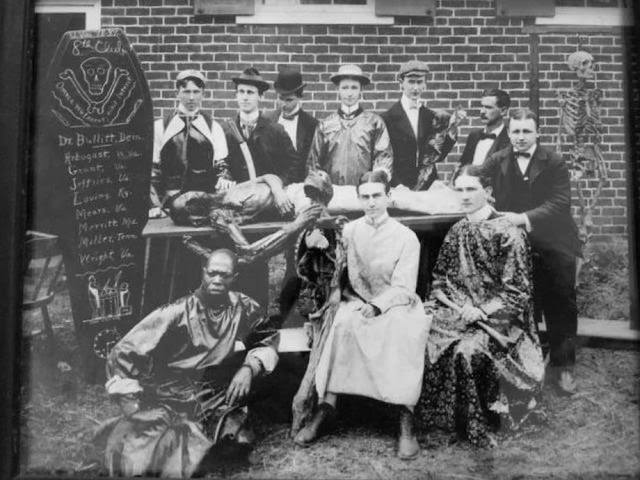
After the Anatomy Act passed, medical schools could operate more openly. Dissection rooms became formal teaching spaces rather than secretive back rooms. Professors structured lessons around specific body systems — the circulatory system one week, the nervous system the next. Students were expected to participate directly, not just observe. Touching, cutting, and naming every structure they encountered was part of the training.
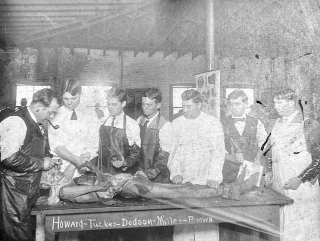
Victorian dissection rooms were nothing like modern anatomy labs. There was no refrigeration. Bodies decomposed quickly, especially in warmer months, which meant sessions moved fast. The smell was overwhelming, and students worked in close quarters with no protective gear beyond an apron. Some accounts describe students eating lunch in the dissection room between lessons — a detail that speaks to how quickly desensitization set in.
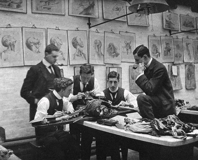
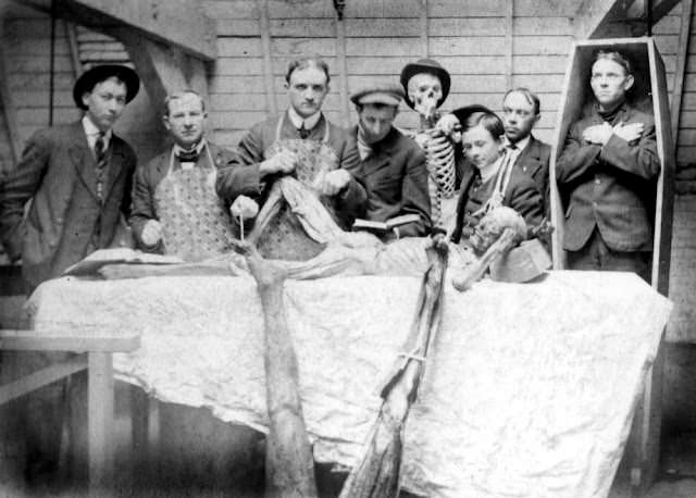
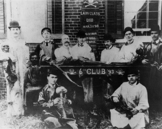
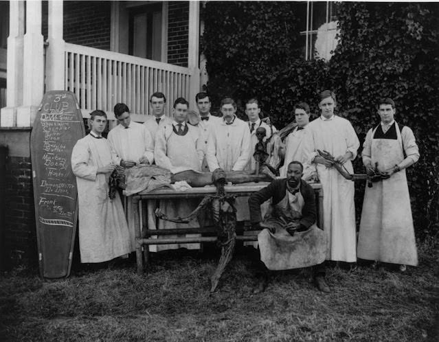
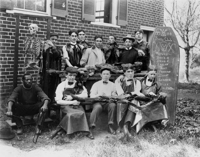
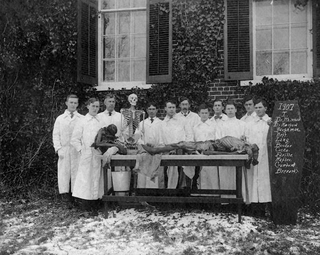
Photography arrived at the perfect historical moment to document this world. The daguerreotype was introduced in 1839, and within a few decades, photographs of medical school life began to appear. Some were formal group portraits taken outside or in lecture halls. Others were taken directly in the dissection room, with students and professors posed around a cadaver. These images were meant to be records of professional achievement — proof that these men had done the work.
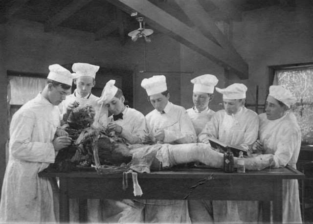
The students in these photographs are almost always men. Women were largely excluded from formal medical education throughout most of the Victorian era. Elizabeth Blackwell became the first woman to earn a medical degree in the United States in 1849, and Elizabeth Garrett Anderson broke the same barrier in England in 1865, but they were exceptions who fought hard against institutional resistance. The dissection rooms in most of these photographs reflect a profession that had not yet opened its doors.
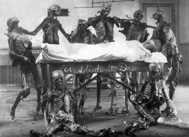
Many of the cadavers in Victorian dissection rooms came from people who had died in poverty. The Anatomy Act’s provision allowing unclaimed bodies from workhouses meant that the poor were far more likely to end up on a dissection table than the wealthy. This created a class dimension to anatomical study that was not lost on critics at the time. For many working-class families, being dissected after death carried a social stigma similar to what execution once carried.
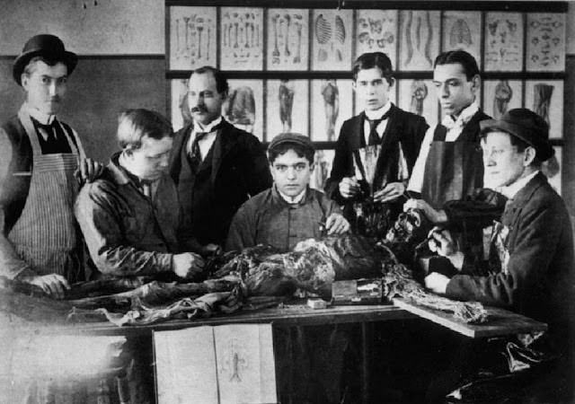
Anatomy professors held enormous authority in Victorian medical schools. The most famous dissection theaters — like those at St. Bartholomew’s Hospital in London or the University of Edinburgh — could seat hundreds of observers. A skilled professor could perform a dissection while lecturing to a packed gallery, naming structures as he cut and expecting students to memorize everything in real time.
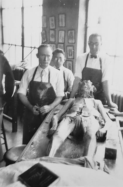
The rise of pathology as a distinct scientific field during this same period deepened the importance of anatomical knowledge. Pathologists studied diseased tissue and organs to understand what had killed a patient, and that work depended entirely on a thorough understanding of what normal anatomy looked like. The dissection table was where both fields overlapped — every anatomy student was also learning to recognize the signs of illness written into the body’s structures.
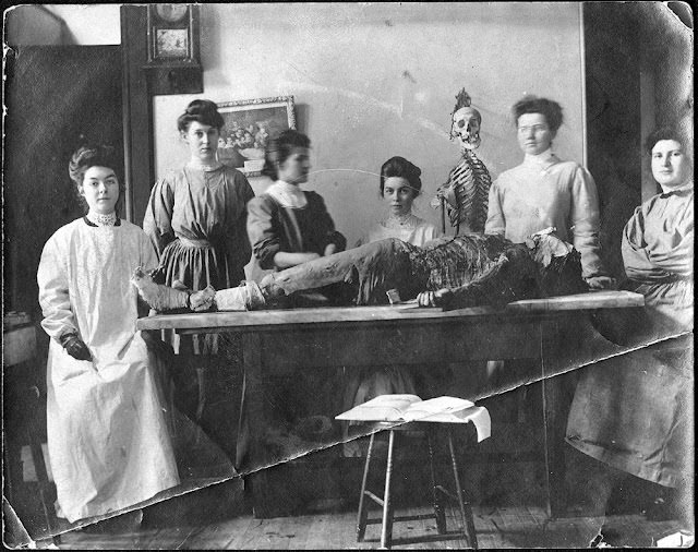
Anatomy atlases and illustrated textbooks flourished during the Victorian era. Publishers produced large, detailed volumes with hand-drawn or engraved plates showing the body layer by layer. Students used them alongside actual dissection, cross-referencing what they saw on the page with what was in front of them on the table. Henry Gray’s “Anatomy: Descriptive and Surgical,” first published in 1858, became the most widely used of these texts and remains in print today.
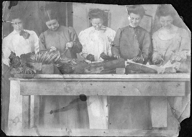
Not every Victorian medical student handled the experience equally. Letters and diaries from the period describe initial shock, nausea, and sleepless nights following early dissection sessions. Some students dropped out. But many accounts also describe a rapid adjustment — within weeks, many students wrote that the work had become routine. The body on the table shifted, in their minds, from a person to a subject of study.
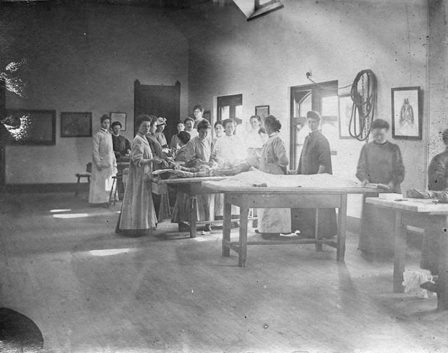
The instruments used in Victorian dissection were simple by modern standards. Scalpels, forceps, bone saws, and probes were the core tools. Students were responsible for caring for their own instruments, sharpening blades and cleaning them between sessions. A well-maintained dissection kit was a point of professional pride. Some surviving kits from the era are housed in medical museums and show the level of craftsmanship that went into their production.
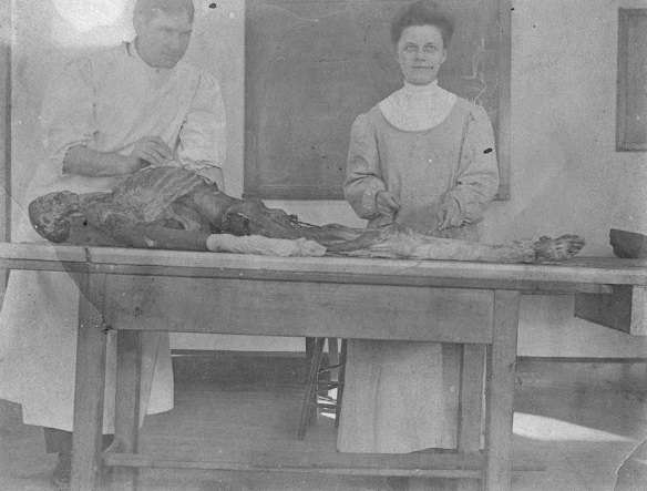
By the end of the 19th century, anatomy education had become more standardized across British and American medical schools. Examinations tested not just a student’s ability to name structures but to understand their function and clinical relevance. The chaotic, legally murky dissection culture of the earlier Victorian period had given way to a regulated system with formal curricula, written examinations, and institutional oversight. The photographs from this era document a profession in the middle of that transformation — raw, hands-on, and unfiltered.
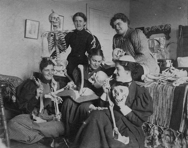

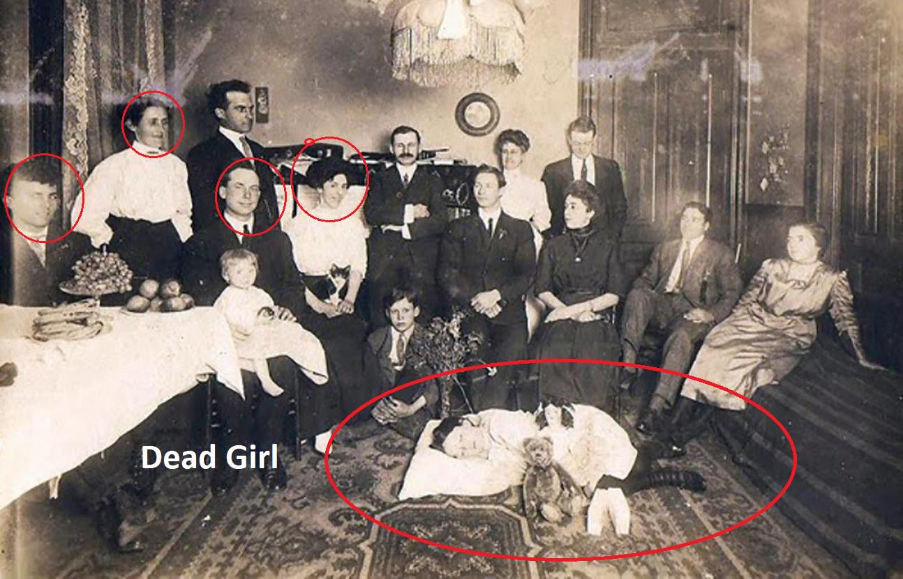
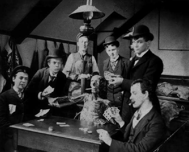
As if that isn’t bad enough… them posing the corpses to take photos with is just really macabre.